. 2009 Jun;14(5):771-81.
doi: 10.1007/s00775-009-0491-y. Epub 2009 Mar 17.

Human serum transferrin: a tale of two lobes. Urea gel and steady state fluorescence analysis of recombinant transferrins as a function of pH, time, and the soluble portion of the transferrin receptor

Affiliations

Human serum transferrin: a tale of two lobes. Urea gel and steady state fluorescence analysis of recombinant transferrins as a function of pH, time, and the soluble portion of the transferrin receptor

Shaina L Byrne et al. J Biol Inorg Chem. 2009 Jun.

Abstract

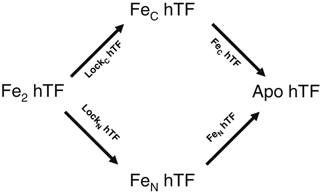
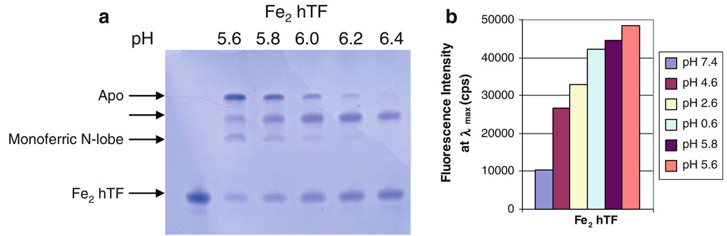
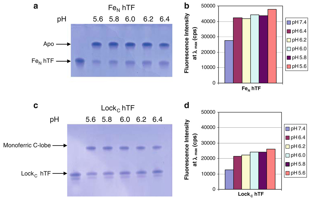
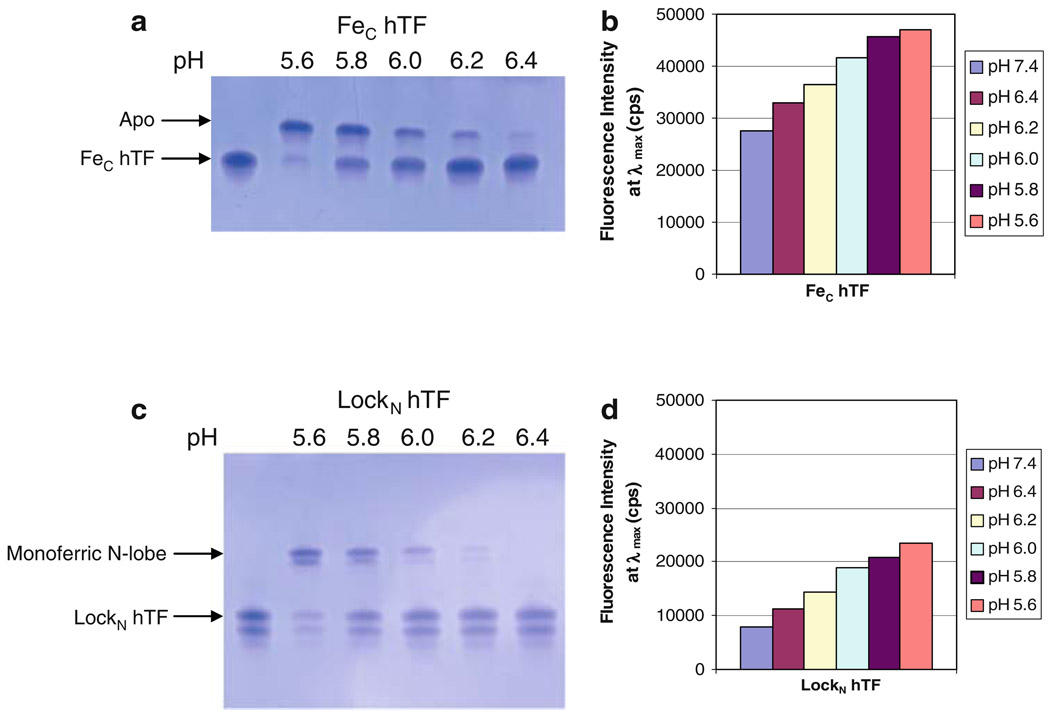
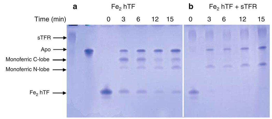
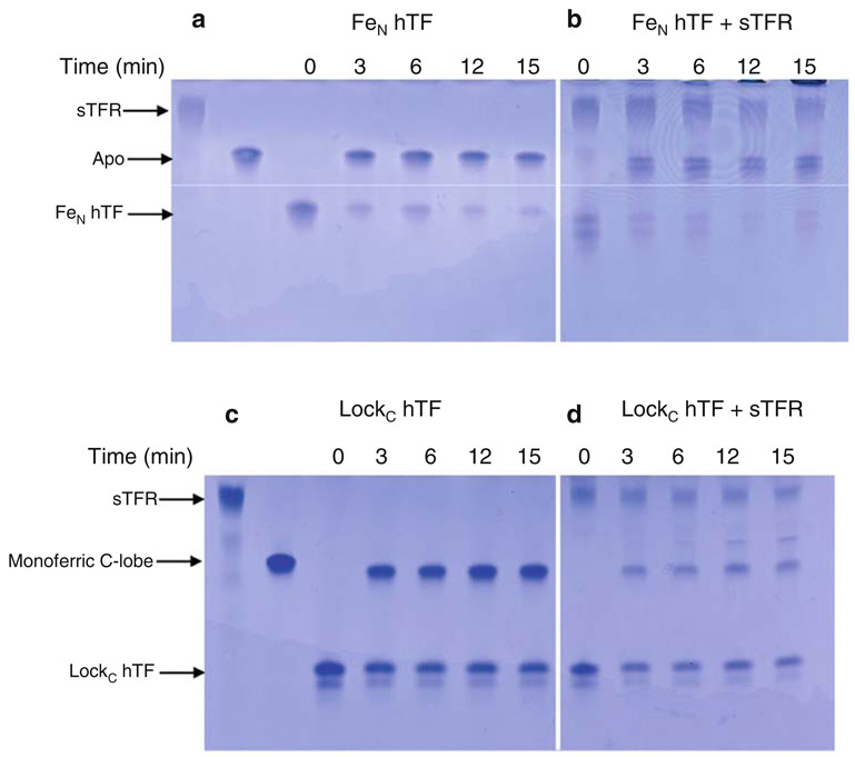
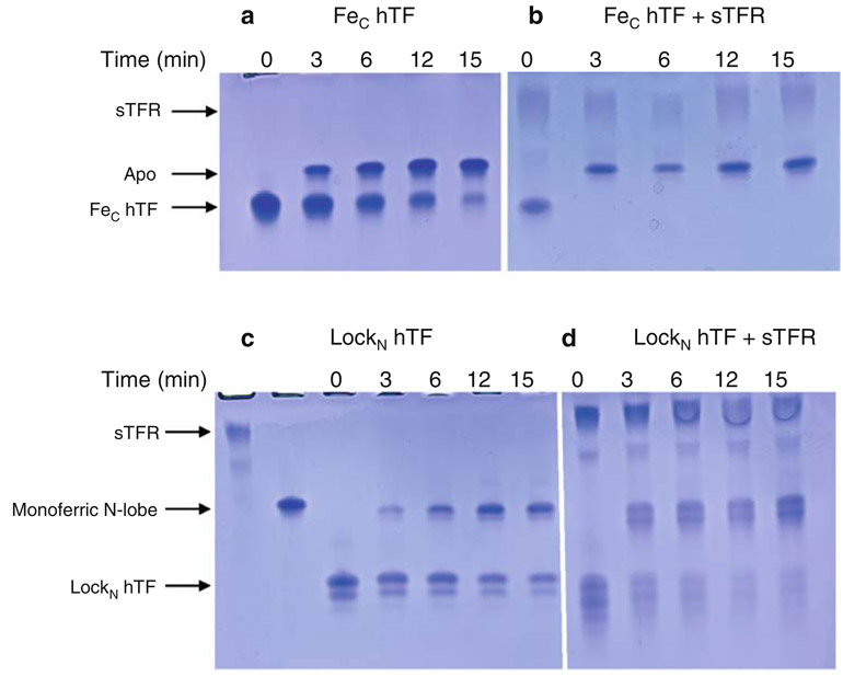
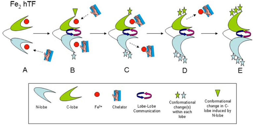
Iron release from human serum transferrin (hTF) has been studied extensively; however, the molecular details of the mechanism(s) remain incomplete. This is in part due to the complexity of this process, which is influenced by lobe-lobe interactions, the transferrin receptor (TFR), the salt effect, the presence of a chelator, and acidification within the endosome, resulting in iron release. The present work brings together many of the concepts and assertions derived from previous studies in a methodical, uniform, and visual manner. Examination of earlier work reveals some uncertainty due to sample and technical limitations. We have used a combination of steady-state fluorescence and urea gels to evaluate the effect of conformation, pH, time, and the soluble portion of the TFR (sTFR) on iron release from each lobe of hTF. The use of authentic recombinant monoferric and locked species removes any possibility of cross-contamination by acquisition of iron. Elimination of detergent by use of the sTFR provides a further technical advantage. We find that iron release from the N-lobe is very sensitive to the conformation of the C-lobe, but is insensitive to the presence of the sTFR or to changes in pH (between 5.6 and 6.4). Specifically, when the cleft of the C-lobe is locked, the urea gels indicate that only about half of the iron is completely removed from the cleft of the N-lobe. Iron release from the C-lobe is most affected by the presence of the sTFR and changes in pH, but is unaffected by the conformation of the N-lobe. A model for iron release from diferric hTF is provided to delineate our findings.


Figures

Fig. 1
Fig. 1
Pathways available to transition from Fe2 hTF to apo hTF. Recombinant constructs used to specifically monitor each route are indicated above/below the arrows
Fig. 2
Fig. 2
Effect of lowering pH on iron release from Fe2 hTF. a 6 M urea gel. All samples were incubated for 15 min in iron-removal buffer before being loaded on the gel (2.5 µg per lane). b Bar graph of steady-state emission intensity at λmax for each spectrum. The emission spectra from which the end points shown in the bar graph were obtained are provided as electronic supplementary material. All samples were incubated for 15 min in iron removal buffer [100 mM MES pH 5.6, 300 mM KCl, 4 mM EDTA], before the emission was monitored. Samples were excited at 280 nm and emission was monitored between 300 and 400 nm using a 320-nm cut-on filter. As an important control, we analyzed a construct with iron locked in both lobes and observed that at pH 5.6 no iron was removed and that the fluorescence intensity was equal to the intensity at pH 7.4, i.e., no iron was removed (data not shown)
Fig. 3
Fig. 3
Effect of lowering pH, and the conformation of the C-lobe, on iron release from the N-lobe of hTF. a 6 M urea gel of FeN hTF, b bar graph of steady-state emission intensity, c 6 M urea gel of LockC hTF, d bar graph of steady-state emission intensity. All samples were prepared as described in the legend to Fig. 2. The appearance of a double band in c is ascribed to the extreme sensitivity of urea gels to charge heterogeneity. We have observed that this heterogeneity seems to increase as a function of the age of the sample and most likely results from oxidation and/or deamination of amino acid side chains
Fig. 4
Fig. 4
Effect of lowering pH, and the conformation of the N-lobe, on iron release from the C-lobe of hTF. a 6 M urea gel of Fec hTF, b bar graph of steady-state emission intensity, c 6 M urea gel of LockN hTF, d bar graph of steady-state emission intensity. All samples were prepared as described in the legend to Fig. 2. See the legend to Fig. 3 for explanation of the appearance of the double band in c in the bands corresponding to the monoferric N-lobe and the diferric species (LockN hTF)
Fig. 5
Fig. 5
Influence of the soluble portion of the transferrin receptor (sTFR) on time-based iron release from Fe2 hTF at pH 5.6. a Fe2 hTF alone, b Fe2 hTF/sTFR complex. All samples were incubated for the designated time courses in 100 mM MES pH 5.6, 300 mM KCl, 4 mM EDTA. Iron release was quenched by the addition of sample buffer
Fig. 6
Fig. 6
Influence of the sTFR on time-based iron release from the N-lobe at pH 5.6. a FeN hTF, b FeN hTF/sTFR complex, c LockC hTF, d LockC hTF/sTFR complex. All samples were incubated for the designated time courses in iron-removal buffer (100 mM MES pH 5.6, 300 mM KCl, 4 mM EDTA). Iron release was quenched by the addition of sample buffer. See the legend to Fig. 3 for explanation of the appearance of the double band in b and c
Fig. 7
Fig. 7
Influence of the sTFR on time-based iron release from the C-lobe at pH 5.6. a FeC hTF, b FeC hTF/sTFR complex, c LockN hTF, d LockN hTF/sTFR complex. All samples were incubated for the designated time courses in 100 mM MES pH 5.6, 300 mM KCl, 4 mM EDTA. Iron release was quenched by the addition of sample buffer. See the legend to Fig. 3 for explanation of the appearance of the double band in c and d
Fig. 8
Fig. 8
Model of iron release from Fe2 hTF. See “Discussion” for full details

References

    1. Halbrooks PJ, He QY, Briggs SK, Everse SJ, Smith VC, Mac-Gillivray RT, Mason AB. Biochemistry. 2003;42:3701–3707. - PubMed
    1. Williams J, Moreton K. Biochem J. 1980;185:483–488. - PMC - PubMed
    1. Huebers H, Josephson B, Huebers E, Csiba E, Finch C. Proc Natl Acad Sci USA. 1981;78:2572–2576. - PMC - PubMed
    1. Mason AB, Halbrooks PJ, James NG, Connolly SA, Larouche JR, Smith VC, MacGillivray RT, Chasteen ND. Biochemistry. 2005;44:8013–8021. - PubMed
    1. He QY, Mason AB. In: Molecular and cellular iron transport. Templeton DM, editor. Toronto: Dekker; 2002.

Publication types