. 2009 Dec;18(10):1493-500.
doi: 10.1089/scd.2009.0052.

Absence of transverse tubules contributes to non-uniform Ca(2+) wavefronts in mouse and human embryonic stem cell-derived cardiomyocytes

Affiliations

Absence of transverse tubules contributes to non-uniform Ca(2+) wavefronts in mouse and human embryonic stem cell-derived cardiomyocytes

Deborah K Lieu et al. Stem Cells Dev. 2009 Dec.

Abstract

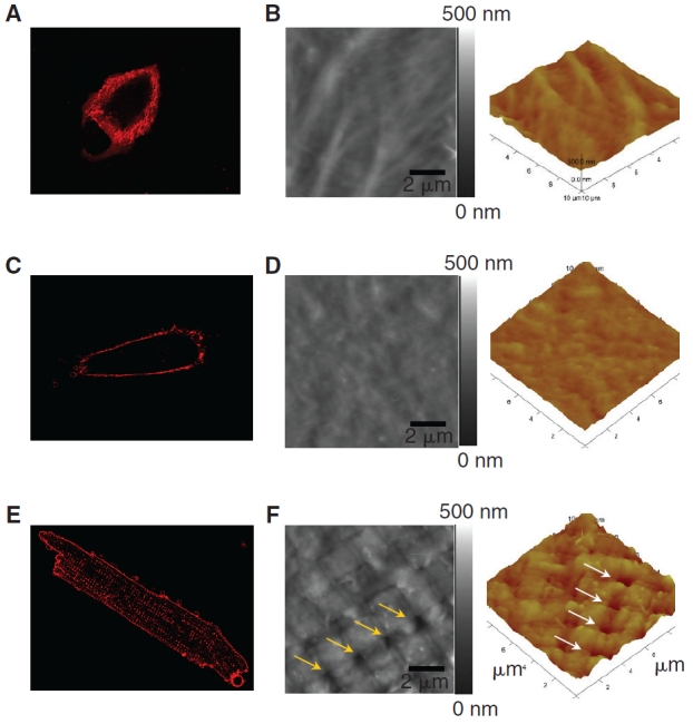
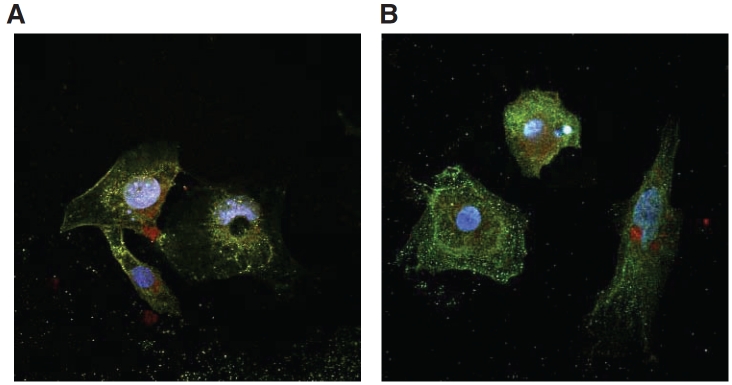
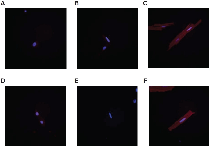
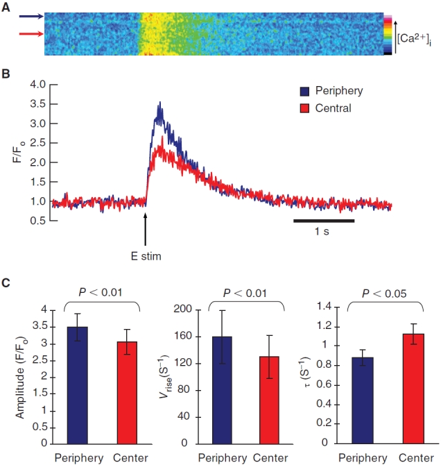
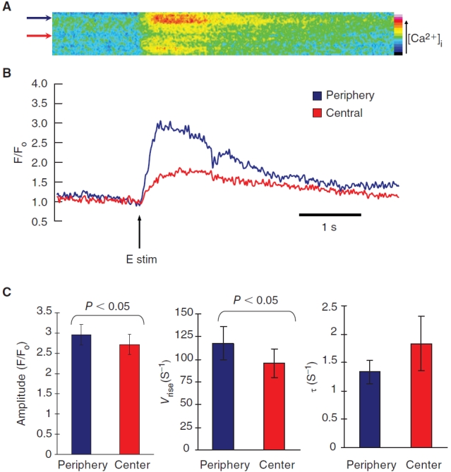
Mouse (m) and human embryonic stem cell-derived cardiomyocytes (hESC-CMs) are known to exhibit immature Ca(2+) dynamics such as small whole-cell peak amplitude and slower kinetics relative to those of adult. In this study, we examined the maturity and efficiency of Ca(2+)-induced Ca(2+) release in m and hESC-CMs, the presence of transverse (t) tubules and its effects on the regional Ca(2+) dynamics. In m and hESC-CMs, fluorescent staining and atomic force microscopy (AFM) were used to detect the presence of t-tubules, caveolin-3, amphiphysin-2 and colocalization of dihydropyridine receptors (DHPRs) and ryanodine receptors (RyRs). To avoid ambiguities, regional electrically-stimulated Ca(2+) dynamics of single ESC-CMs, rather than spontaneously beating clusters, were measured using confocal microscopy. m and hESC-CMs showed absence of dyads, with neither t-tubules nor colocalization of DHPRs and RyRs. Caveolin-3 and amphiphysin-2, crucial for the biogenesis of t-tubules with robust expression in adult CMs, were also absent. Single m and hESC-CMs displayed non-uniform Ca(2+) dynamics across the cell that is typical of CMs deficient of t-tubules. Local Ca(2+) transients exhibited greater peak amplitude at the peripheral than at the central region for m (3.50 +/- 0.42 vs. 3.05 +/- 0.38) and hESC-CMs (2.96 +/- 0.25 vs. 2.72 +/- 0.25). Kinetically, both the rates of rise to peak amplitude and transient decay were faster for the peripheral relative to the central region. Immature m and hESC-CMs display unsynchronized Ca(2+) transients due to the absence of t-tubules and gene products crucial for their biogenesis. Our results provide insights for driving the maturation of ESC-CMs.


Figures

FIG. 1.
FIG. 1.
T-tubule imaging of a mouse embryonic stem cell-derived cardiomyocyte (mESC-CM), a human ESC-CM (hESC-CM), and a mature ventricular CM. Di-8-ANEPPS confocal microscopic images of an mESC-CM (A) and an hESC-CM (C) did not show intracellular fluorescent spots like those in an adult guinea pig ventricular CM (E), suggesting the absence of t-tubules. The absence of t-tubules in ESC-CMs was further confirmed by AFM imaging of an adult guinea pig ventricular cardiomyocyte (F), showing regularly spaced pores in the sarcolemma that coincide with the z-lines, while mESC-CM (B) and hESC-CM (D) surface showed comparatively smoother topology with no presence of invaginations that are indicative of t-tubules.
FIG. 2.
FIG. 2.
Double immunostaining of DHPRs and RyRs in mouse and human embryonic stem cell-derived cardiomyocytes (mESC-CMs and hESC-CMs). DHPRs and RyRs showed lack of colocalization in mESC-CMs (A) and hESC-CMs (B). All cells were nuclear counterstained with Hoechst 33342.
FIG. 3.
FIG. 3.
Immunostaining of caveolin-3 and amphiphysin-2 in mouse embryonic stem cell-derived cardiomyocytes (mESC-CMs), human ESC-CMs (hESC-CMs), and mature adult CMs. Caveolin-3 was absent in both mESC-CMs (A) and hESC-CMs (B). Adult ventricular CMs were stained as positive control (C). Amphiphysin-2 was also deficient in mESC-CMs (D) and hESC-CMs (E). Amphiphysin-2 was also stained in adult ventricular CMs as positive control (F). All cell nuclei were counterstained with Hoechst 33342.
FIG. 4.
FIG. 4.
Electrically-induced transient Ca2+ increase in mouse embryonic stem cell-derived cardiomyocytes (mESC-CMs). (A) Time progression linescans of pseudo-colored transient increase in intracellular Ca2+ across the midplane of a mESC-CM showed a U-shaped wavefront typical of CMs lacking t-tubules. (B) Quantified Ca2+ transient of linescans in A. The arrow indicates time of pulsed electrical stimulus. (C) The peak amplitude increase was statistically greater for the periphery relative to the center (n = 12). The transient rate of rise (V rise) and the transient decay as shown by the decay time constant (τ) were both statistically faster at the peripheral than the central region. Data shown as mean ± SEM.
FIG. 5.
FIG. 5.
Electrically-induced transient Ca2+ increase in adult guinea pig ventricular cardiomyocytes (CMs) and coupled cluster of mouse embryonic stem cell-derived CMs (mESC-CMs). (A) Time progression linescans show uniform Ca2+ wavefronts in mature adult ventricular CMs typical of CMs with t-tubules. (B) Time progression linescans of mESC-CMs in coupled cluster exhibited the same U-shaped Ca2+ wavefronts as the single mESC-CMs. The arrows indicate time of electrical stimulation.
FIG. 6.
FIG. 6.
Electrically-induced transient Ca2+ increase in human embryonic stem cell-derived cardiomyocytes (hESC-CMs). (A) Time progression linescans of pseudo-colored transient increase in intracellular Ca2+ across the midplane of a hESC-CM showed a U-shaped wavefront typical of CMs lacking t-tubules. (B) Quantified Ca2+ transient of linescans in A. The arrow indicates time of pulsed electrical stimulus. (C) The peak amplitude increase was statistically greater for the periphery relative to the center (n = 11). The transient rate of rise (V rise) was statistically faster at the periphery than the central region. The transient decay was faster for the periphery as shown by their decay time constant (τ), but the difference did not reach statistical significance. Data shown as mean ± SEM.

References

    1. Liu J, Fu JD, Siu CW, Li RA. Functional sarcoplasmic reticulum for calcium-handling of human embryonic stem cell-derived cardiomyocytes: insights for driven maturation. Stem Cells. (2007);25:3038–3044. - PubMed
    1. Sartiani L, Bettiol E, Stillitano F, Mugelli A, Cerbai E, Jaconi ME. Developmental changes in cardiomyocytes differentiated from human embryonic stem cells: a molecular and electrophysiological approach. Stem Cells. (2007);25:1136–1144. - PubMed
    1. Itzhaki I, Schiller J, Beyar R, Satin J, Gepstein L. Calcium handling in embryonic stem cell-derived cardiac myocytes: of mice and men. Ann N Y Acad Sci. (2006);1080:207–215. - PubMed
    1. Bers DM. Cardiac excitation–contraction coupling. Nature. (2002);415:198–205. - PubMed
    1. Brette F, Orchard C. T-tubule function in mammalian cardiac myocytes. Circ Res. (2003);92:1182–1192. - PubMed

Publication types

LinkOut - more resources