

Review
doi: 10.1111/j.1600-065X.2008.00760.x.
SHP-1 and SHP-2 in T cells: two phosphatases functioning at many levels
Affiliations
- PMID: 19290938
- PMCID: PMC2669678
- DOI: 10.1111/j.1600-065X.2008.00760.x
Item in Clipboard
Review
SHP-1 and SHP-2 in T cells: two phosphatases functioning at many levels
Immunol Rev.
2009 Mar.
Abstract
Tyrosine phosphorylation and dephosphorylation of proteins play a critical role for many T-cell functions. The opposing actions of protein tyrosine kinases (PTKs) and protein tyrosine phosphatases (PTPs) determine the level of tyrosine phosphorylation at any time. It is well accepted that PTKs are essential during T-cell signaling; however, the role and importance of PTPs are much less known and appreciated. Both transmembrane and cytoplasmic tyrosine phosphatases have been identified in T cells and shown to regulate T-cell responses. This review focuses on the roles of the two cytoplasmic PTPs, the Src-homology 2 domain (SH2)-containing SHP-1 and SHP-2, in T-cell signaling, development, differentiation, and function.
Figures
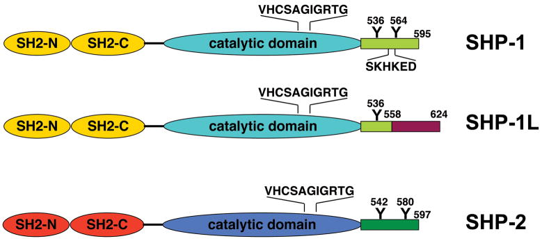
Defined domain within the proteins are indicated. Moreover, the PTP signature sequence, potential tyrosine phosphorylation sites, and the lipid rafts targeting peptide are marked. The differences in the C-termini between SHP-1 and SHP-1L are shown.
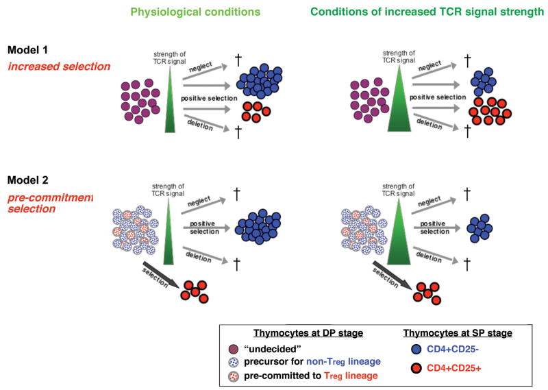
In the model of increased selection (model 1), thymocytes undergoing the selection process at the DP stage are ‘undecided’ and can develop into Treg or conventional non-Treg cells depending on the strength of signal they encounter. CD4+CD25+ Treg cells are selected in response to a defined range of TCR signaling strength that falls between positive selection of non-Treg cells and negative selection/deletion (left panel). Under conditions of increased TCR signaling strength (right panel), such as exposure of the TCR to a high affinity ligand or loss of the negative regulator SHP-1, the selection process shifts towards positive selection of CD4+CD25+ Treg cells and greater deletion of non-Treg cells, resulting in an increase in the absolute number of CD4+CD25+ T cells. In contrast, in the model of pre-commitment/selection (model 2), a defined fraction of the CD4+CD8+ DP thymocytes is already committed to the CD4+CD25+ Treg cell lineage. Whether the pre-commitment is driven by encounter with a signal or by a stochastic process is unknown at this time. The remaining non-committed DP cells have the potential to develop into conventional non-Treg T cells upon passing of positive/negative selection, but lost the potential to develop into Treg cells. The cells pre-committed to the Treg lineage still require additional signals and might undergo a selection process similar to conventional T cell to acquire the Treg phenotype but their absolute numbers are unaffected by the TCR signal strength as long as it is above a certain threshold. Moreover, even when exposed to strong TCR-mediated signaling, these developing CD4+CD25+ Treg cells are highly resistant to deletion. Therefore under conditions of strong TCR signaling strength (right panel), the observed gain in the percentage of CD4+CD25+ Treg cells is due to increased negative selection and selective loss of the CD4+CD25- conventional T cell population.
References
-
- Yarden Y, Ullrich A. Growth factor receptor tyrosine kinases. Ann Rev Biochem. 1988;57:443–478. - PubMed
-
- Hunter T. Protein modification: phosphorylation on tyrosine residues. Curr Opin Cell Biol. 1989;1:1168–1181. - PubMed
-
- Ullrich A, Schlessinger J. Signal transduction by receptors with tyrosine kinase activity. Cell. 1990;61:203–212. - PubMed
-
- Alonso A, Sasin J, Bottini N, Friedberg I, Osterman A, Godzik A, Hunter T, Dixon J, Mustelin T. Protein tyrosine phosphatases in the human genome. Cell. 2004;117:699–711. - PubMed
Publication types
MeSH terms
Substances
Grants and funding
LinkOut - more resources
Full Text Sources
Other Literature Sources
Miscellaneous
