. 2009 Mar 16:4:11.
doi: 10.1186/1749-8104-4-11.

Elevated P75NTR expression causes death of engrailed-deficient midbrain dopaminergic neurons by Erk1/2 suppression

Affiliations

Elevated P75NTR expression causes death of engrailed-deficient midbrain dopaminergic neurons by Erk1/2 suppression

Kambiz N Alavian et al. Neural Dev. .

Abstract

Background: The homeodomain transcription factors Engrailed-1 and Engrailed-2 are required for the survival of mesencephalic dopaminergic (mesDA) neurons in a cell-autonomous and gene-dose-dependent manner. Homozygote mutant mice, deficient of both genes (En1-/-;En2-/-), die at birth and exhibit a loss of all mesDA neurons by mid-gestation. In heterozygote animals (En1+/-;En2-/-), which are viable and fertile, postnatal maintenance of the nigrostriatal dopaminergic system is afflicted, leading to a progressive degeneration specific to this subpopulation and Parkinson's disease-like molecular and behavioral deficits.

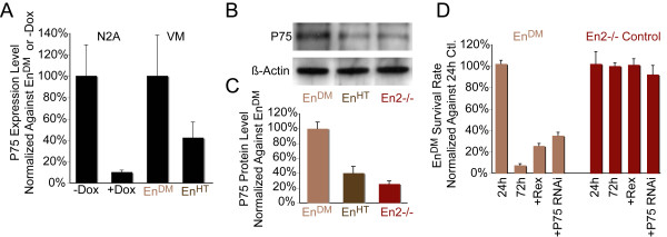
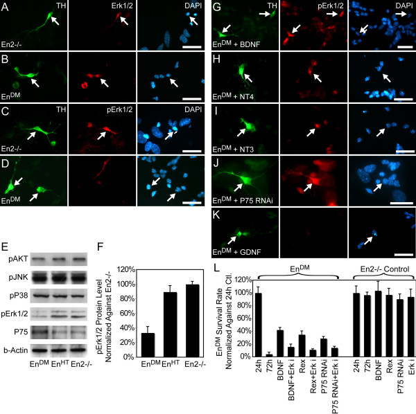
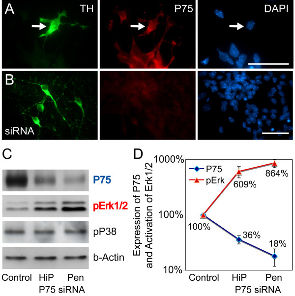
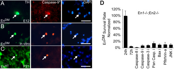
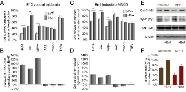
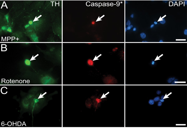
Results: In this work, we show that the dose of Engrailed is inversely correlated to the expression level of the pan-neurotrophin receptor gene P75NTR (Ngfr). Loss of mesDA neurons in the Engrailed-null mutant embryos is caused by elevated expression of this neurotrophin receptor: Unusually, in this case, the cell death signal of P75NTR is mediated by suppression of Erk1/2 (extracellular-signal-regulated kinase 1/2) activity. The reduction in expression of Engrailed, possibly related to the higher levels of P75NTR, also decreases mitochondrial stability. In particular, the dose of Engrailed determines the sensitivity to cell death induced by the classic Parkinson-model toxin MPTP and to inhibition of the anti-apoptotic members of the Bcl-2 family of proteins.

Conclusion: Our study links the survival function of the Engrailed genes in developing mesDA neurons to the regulation of P75NTR and the sensitivity of these neurons to mitochondrial insult. The similarities to the disease etiology in combination with the nigral phenotype of En1+/-;En2-/- mice suggests that haplotype variations in the Engrailed genes and/or P75NTR that alter their expression levels could, in part, determine susceptibility to Parkinson's disease.


Figures

Figure 1
Figure 1
Elevated P75NTR expression is causal for cell death. (A) Quantitative RT-PCR of ventral midbrain tissue (VM) derived from EnDM (En1-/-;En2-/-) and EnHT E12 embryos, and of En1-expressing N2A cells inducible by doxycycline (Dox). P75NTR expression is inversely correlated with En1 expression levels in tissues and cell lines. (B, C) Western blot analysis of the ventral midbrain tissue shows the same relationship between P75NTR protein levels and En1 expression. Each active En1 allele decreases the P75NTR expression level (n = 3, p = 0.002). (D) Ventral midbrain cultures derived from EnDM and En2-/- embryos. Silencing of P75NTR by double-stranded RNA oligos and application of P75NTR-inhibiting antibody (Rex) increases the survival rate of EnDM mesDA neurons compared to untreated control (Ctl) or after treatment with scrambled RNA oligos (n ≥ 6, p < 0.01). Error bars indicate standard error.
Figure 2
Figure 2
Loss of Engrailed induces neurotrophin requirement in mesDA neurons. (A-F) Double immunohistochemistry on dissociated cells derived from En2-/- (A-C) and En1-/-;En2-/- (EnDM) (D-F) E12 ventral midbrain using antibodies against tyrosine kinase (Trk)B (A, D), TrkC (B, E), P75NTR(C, F) and TH (green) counterstained with DAPI. TrkB, TrkC and P75NTR are expressed by TH+ cells from both genotypes; however, the immunohistochemistry is not sensitive enough to detect differences in P75NTR expression between genotypes. (G, H) Western blot of ventral midbrain tissue derived from different Engrailed genotypes. The two Trk receptors do not depend on Engrailed expression (G). Brain-derived neurotrophic factor (BDNF), neurotrophin (NT)4 and NT3 are not expressed in E12 ventral midbrain tissue, but they are in the adult (H). (I) Treatments (>10 ng/ml) for 72 hours with TrkB/C-specific neurotrophins – BDNF, NT4 and NT3 – greatly increases the survival rate of EnDM mesDA neurons (n ≥ 6; p < 0.001), whereas nerve growth factor (NGF), glial cell line-derived neurotrophic factor (GDNF), transforming growth factor (TGF)-β and growth differentiation factor (GDF)-15 do not significantly alter survival rate. (J) Dose response curve: BDNF concentration plotted against survival rate showing saturation at approximately the 10 ng/ml. Scale bars: 25 μm. Error bars indicate standard error. Ctl, control.
Figure 3
Figure 3
Differential activation of Erk1/2 in mesDA neurons. (A-D, G-K) Immunohistochemistry of E12 ventral midbrain cell culture stained against TH (green), total Erk1/2 protein (red) (A, B) and phosphorylated Erk1/2 (red) (C-D, G-K). (A-D) While Erk1/2 protein is present in mesDA neurons of both genotypes (A, B), it is only phosphorylated in En2-/- mesDA neurons (C) and not in the En1-/-;En2-/- (EnDM) counterparts (D). (E-I) Erk1/2 becomes activated in EnDM mesDA neurons after treatment with the survival-inducing neurotrophins, brain-derived neurotrophic factor (BDNF), neurotrophin (NT)4 and NT3, or after silencing of P75NTR (RNA interference (RNAi)) (G-J), but not when glial cell line-derived neurotrophic factor (GDNF) is applied (I). (E) Western blot of E12 ventral midbrain tissue confirms the immunohistochemical finding of differential phosphorylation between genotypes and shows that neither AKT, part of the phosphotidyl inositol-3 kinase pathway, nor other mitogen-activated protein kinases, such as JNK and P38, are differentially activated. (F) Quantification of phosphorylated Erk1/2 in western blot normalized against En2-/- tissue. (L) Number of TH-positive cells in EnDM and En2-/- ventral midbrain cultures after 72 hours, treated with the 400 nM Mek inhibitor U0126 in conjunction with BDNF, Penetratin-coupled P75NTR double-stranded RNA oligonucleotides and the P75NTR inhibiting antibody (Rex). Numbers are normalized against untreated cultures at 24 hours. The rescue effect is significantly reduced when the EnDM cultures are treated with the Erk1/2 inhibitor. Scale bars: 25 μm. Error bars indicate standard error. Ctl, control.
Figure 4
Figure 4
Erk1/2 activation is inversely correlated with P75NTR expression. (A, B) Immunohistochemistry on E12 dissociated ventral midbrain cultures derived from EnHT E12 embryos stained against TH (green) and P75NTR. P75NTR expression is absent in cultures treated with Penetratin-coupled double-stranded RNA oligos. (C) Western blot of ventral midbrain cultures using two methods of RNA transfection with different efficiencies: HiPerfect (HiP) and Penetratin-coupled RNA oligos show increase in phosphorylated Erk1/2 after silencing of P75NTR, but no changes in phosphorylation of P38. (D) Quantification of P75NTR expression and Erk1/2 phosphorylation after transfection with P75NTR double-stranded RNA oligonucleotides, normalized against untreated EnHT cultures, showing the inverse correlation between the two parameters. Scale bars: 25 μm. Error bars indicate standard error.
Figure 5
Figure 5
Mitochondrial apoptosis in Engrailed-deficient mesDA neurons. (A-C) TH and activated caspase-9 immunohistochemistry on ventral midbrain coronal sections of E13 En1-/-;En2-/- (EnDM) embryos (A), of En1+/-;En2-/- (EnHT) postnatal day 20 brain (C), and of EnDM E12 cell culture (B) counterstained with the nuclear marker DAPI. Dying TH-positive neurons (arrows) exhibit small rounded cell bodies and signs of apoptosis, that is, activated caspase-9 and pyknotic nuclei (DAPI). (D) EnDM ventral midbrain cultures treated for 72 hours with inhibitors for caspases-3, -8 and -9, a pan-caspase inhibitor, z-vad-fmk, the BAX inhibitor Ku70, the P53 inhibitor Pfithrin-α, and the JNK inhibitor SP600125. None of the treatments significantly changed the survival rate of EnDM mesDA neurons. The number of surviving TH-positive cells was normalized in each case against untreated cultures 24 hours after dissociation. Scale bars: 25 μm. Error bars indicate standard error.
Figure 6
Figure 6
The sensitivity to induction of the intrinsic pathway of apoptosis correlates with En1 expression. (A-D) Ventral midbrain cultures 24 hours after application of apoptosis-inducing compounds. Charts of EnHT (En1+/-;En2-/-) and En2-/- E12 cultures depicting the number of surviving TH-positive cells (A, B) and cultures of En1-inducible MN9D cells depicting cell survival measured by cell proliferation assay (C, D). Surviving cells were normalized against untreated EnHT cultures (A) or untreated non-induced MN9D cells (C), treated EnHT cultures (B) or treated non-induced MN9D cells (D). Higher Engrailed expression reduced the cell death rate after MPP+, HA14-1 and chelerythrine chloride (CC) treatment, whereas the rate of cell survival after application of the tumor necrosis factor alpha (TNFa), Prima-1 and Apoptosis Activator-2 (AA2) does not correlate with the level of En1 expression. Dox, doxycycline. (E) Western blot analysis of mitochondrial and cytoplasmic protein fractions of MN9D cells 72 hours after En1 induction and 24 hours after MPP+ treatment. (F) Proportion of cytochrome C (Cyt-C) in cytosol is lower in En1-expressing MN9D cells before and after MPP+ treatment. Scale bars: 25 μm. Error bars indicate standard error. 6 ≤ n ≤ 27, *p ≤ 0.001, **p ≤ 0.0001. Cyt, cytosolic; Mit, mitochondrial; ANT, adenine nucleotide transporter; Dox, doxycycline.
Figure 7
Figure 7
Mitochondrial apoptosis after complex I inhibition. (A-C) Immunohistochemistry of E12 ventral midbrain cell cultures. Caspase-9 is activated in wild-type mesDA neurons (arrows), treated with MPP+, rotenone, or 6-OHDA.

Similar articles

Cited by

References

    1. Schultz W. Getting formal with dopamine and reward. Neuron. 2002;36:241–263. doi: 10.1016/S0896-6273(02)00967-4. - DOI - PubMed
    1. Girault JA, Greengard P. The neurobiology of dopamine signaling. Arch Neurol. 2004;61:641–644. doi: 10.1001/archneur.61.5.641. - DOI - PubMed
    1. Abou-Sleiman PM, Muqit MM, Wood NW. Expanding insights of mitochondrial dysfunction in Parkinson's disease. Nat Rev Neurosci. 2006;7:207–219. doi: 10.1038/nrn1868. - DOI - PubMed
    1. Tatton WG, Chalmers-Redman R, Brown D, Tatton N. Apoptosis in Parkinson's disease: signals for neuronal degradation. Ann Neurol. 2003;53:S61–70. doi: 10.1002/ana.10489. discussion S70-62. - DOI - PubMed
    1. Farrer MJ. Genetics of Parkinson disease: paradigm shifts and future prospects. Nat Rev Genet. 2006;7:306–318. doi: 10.1038/nrg1831. - DOI - PubMed

Publication types

MeSH terms

LinkOut - more resources