. 2009 Mar 16:9:22.
doi: 10.1186/1471-213X-9-22.

Expression of transmembrane carbonic anhydrases, CAIX and CAXII, in human development

Affiliations

Expression of transmembrane carbonic anhydrases, CAIX and CAXII, in human development

Shu-Yuan Liao et al. BMC Dev Biol. .

Abstract

Background: Transmembrane CAIX and CAXII are members of the alpha carbonic anhydrase (CA) family. They play a crucial role in differentiation, proliferation, and pH regulation. Expression of CAIX and CAXII proteins in tumor tissues is primarily induced by hypoxia and this is particularly true for CAIX, which is regulated by the transcription factor, hypoxia inducible factor-1 (HIF-1). Their distributions in normal adult human tissues are restricted to highly specialized cells that are not always hypoxic. The human fetus exists in a relatively hypoxic environment. We examined expression of CAIX, CAXII and HIF-1alpha in the developing human fetus and postnatal tissues to determine whether expression of CAIX and CAXII is exclusively regulated by HIF-1.

Results: The co-localization of CAIX and HIF-1alpha was limited to certain cell types in embryonic and early fetal tissues. Those cells comprised the primitive mesenchyma or involved chondrogenesis and skin development. Transient CAIX expression was limited to immature tissues of mesodermal origin and the skin and ependymal cells. The only tissues that persistently expressed CAIX protein were coelomic epithelium (mesothelium) and its remnants, the epithelium of the stomach and biliary tree, glands and crypt cells of duodenum and small intestine, and the cells located at those sites previously identified as harboring adult stem cells in, for example, the skin and large intestine. In many instances co-localization of CAIX and HIF-1alpha was not evident. CAXII expression is restricted to cells involved in secretion and water absorption such as parietal cells of the stomach, acinar cells of the salivary glands and pancreas, epithelium of the large intestine, and renal tubules. Co-localization of CAXII with CAIX or HIF-1alpha was not observed.

Conclusion: The study has showed that: 1) HIF-1alpha and CAIX expression co- localized in many, but not all, of the embryonic and early fetal tissues; 2) There is no evidence of co-localization of CAIX and CAXII; 3) CAIX and CAXII expression is closely related to cell origin and secretory activity involving proton transport, respectively. The intriguing finding of rare CAIX-expressing cells in those sites corresponding to stem cell niches requires further investigation.


Figures
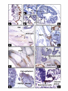
Figure 1
Figure 1
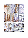
The embryonic period (4–8 weeks): CAIX expression was first seen in the cytotrophoblasts (A, arrow) and certain cells in the embryo (B), such as primitive mesenchyma (B1 long arrow), external surface of the embryo (B2, short double arrows), and mesenchymal cells of the chorion (B2, double long arrows). In the later embryonic stage, CAIX expression was primarily observed in the mesencyhmal cells involving chondrogenesis, as shown in the facial bone (C), bronchial tree (D), the limb (E), and the pelvic bone (F). All of the epithelial cells lining the body cavity were also positive for CAIX, e.g. the peritoneum (F), and the surface of the gonad (G). The only CAIX-positive cells in the CNS are ependymal cells (H). Original magnifications: A, B and H (20×); B1 and B2 (40×); C, D and F (4×); E and G (10×).
Figure 2
Figure 2
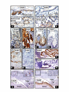
CAIX expression in the placenta, skin, squamous mucosa and skeletal system. The mesenchymal cells of the chorionic plate and umbilical cord retained their CAIX expression until birth (A1, A2). In the epidermis, CAIX expression was limited to the basal layer (B1, F), hair buds (B1) and sebaceous units, hair follicles and the bulges (B2, arrow and inserts). Between 18 to 24 months after birth, CAIX expression was restricted to the hair follicles, sebaceous units, and the infundibulum (C). In the skeletal system, persistent high levels of CAIX immunoreactivity was seen in the chondrocytes, mesenchymal condense (E, F, G) and tendoligamental tissues (G, H). The basal layer of squamous mucosa of the nose (D) and oral cavity (G) also expressed CAIX. W = gestational age in weeks; M = postnatal age in months. Original magnifications: A1, A2, B, C and H (20×); D and G (4×); E (10×) and F (40×).
Figure 3
Figure 3
CAIX expression in bronchial trees and genital organs: In the bronchial trees high expression of CAIX was seen in the peribronchial immature mesenchymal cells and cartilage (designated C) (A1) but progressively diminished when the tissues became mature (A2, A3). CAIX positivity was persistently seen in peritoneal lining cells as shown in B (insert) and G1, G2 (surface epithelium [SE], arrow). As early as the 13th week of gestation, CAIX positive cells were seen in the flat SE of the ovary, the outer muscular layer and rare epithelial cells of the uterus and fallopian tubes (B). Around 26 to 27 weeks, high levels of CAIX expression were transiently seen in the epithelium of fallopian tubes (C1), endometrium (D1), and the cervix (E1). After birth, there was either no CAIX expression (C3) or expression was limited to occasional endometrial cells (D2), reserve cells of the cervix (E2, and corresponding H&E stain in insert, and F1, F2). In the ovary, CAIX expression was observed in SE migrating into the stroma and forming the primordial follicle (PF) (G1, G2). Persistent expression of CAIX was observed in the coelomic remnants: rete testis (H1) and tubule reti (H2). W = gestational age in weeks; D, M = postnatal age in days and months, respectively. Original magnifications: A1, A2, A3, C1, C2, C3, E1, E2 and F2 (20×); B (4×); H1 and H2 (10×); D1, D2, F1, G1 and G2 (40×).
Figure 4
Figure 4
In the adrenal gland (A), strong CAIX immunoreactivity was seen in many cortical cells near the capsule (17W), but the level of expression progressively diminished after birth and, by the end of two years, no CAIX positive cells were seen. In the thymus gland (B), CAIX positive cells were primarily identified near the Hassall's corpuscles (16W) but disappeared by the first year after birth (24M). In the stomach (C), CAIX expression was first seen in surface columnar cells at the 9th week of gestation (9W). At 17 weeks, diffuse immunoreactivity was seen in the glandular and surface columnar cells (17W). This level of expression persisted after birth (24M). In the small intestine CAIX positive cells were restricted to the crypts (D1) and in the large intestine only rare positive cells were identified near the base of the crypts (D2). In the biliary trees, CAIX expression was seen in the epithelium of the entire ductal system and was persistent after birth. The liver (E1) and pancreas (E2) are shown as examples. During early fetal period, extensive CAIX expression was seen in the basal/reserve cells of the respiratory epithelium (F1, 14W) but after birth only rare reserve cells continued to express CAIX (F2, 24M). W = gestational age in weeks; D, M = postnatal age in days and months, respectively. Original magnifications: A1, A2, A3, B1 and C (20×); B2, D1, D2, E1, E2, F1 and F2 (40×).
Figure 5
Figure 5
Schema of CAIX expression in the cytotrophoblast and extra-/intra-embryonic mesoderm derivatives during human development (embryonic, fetal and postnatal periods).
Figure 6
Figure 6
Schema of CAIX expression in the ectodermal and endodermal derivatives during human development (embryonic, fetal and postnatal periods).
Figure 7
Figure 7
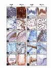
Comparison of CAIX and CAXII expression during human development. There is no correlation between CAXII and CAIX expression. CAXII positive cells were distributed in taste buds and the submucosal gland of the tongue (A, 17W), the acinar cells of the pancreas (B, lower panel, but not ductal cells [B, upper panel]), the parietal cells but not columnar cells of the stomach (C1, 27W), the large intestine (D1, 17W), the distal tubules and collecting ducts of the kidney (E1, 8W), the choroid plexus (F1, 8W) and the remnants of mesonephric ducts of the ovary (G, 13W). In contrast, CAIX positive cells were seen in the gastric columnar cells (C2, 27W), the ependymal cells (F2, 8W), and the surface epithelial cells of the ovary and peritoneum (H, 13W). However, CAIX expression was not seen in the large intestine (D2, 17W), kidney (E2, 8W) and the remnants of the ovarian mesonephric ducts (H, 13W). W = gestational age in weeks. Original magnifications: A, F1 and F2 (20×); B, C1, C2, D1 and D2 (40×); E1, E2, G and H (10×).
Figure 8
Figure 8
The correlation between CAIX and HIF-1α expression during human development. In the early stage (5–9 weeks of gestation) diffuse HIF-1α immunoreactivity was seen in the CNS (A), primitive intestine (B), chorionic villi (C) and chondrocytes of the disc (D), mesenchymal cells and chondrocytes of the limb (E), and the skin (E arrow). In contrast, no CAIX expression was seen in the CNS (A), primitive intestine (B), and chorionic villi (C). Co-expression of HIF-1α and CAIX was seen in the skin (E) and the cells involved in chondrogenesis (D, E). During later human development, around 19–20 weeks, HIF-1α expression was persistently observed in the kidney (F) and rectum (G) but no CAIX expressing cells were seen in these organs. However, a degree of overlap between CAIX and HIF-1α expression was seen in the skin (H), squamous mucosa of the vagina and the cervix (I and J). W = gestational age in weeks. Original magnifications: A, B, D-H (40×); C (left panel 20×; right panel 40×); I (20×); J (left panel 20×; right panel 40×).

References

    1. Supuran CT. Carbonic anhydrases-an overview. Curr Pharm Des. 2008;14:603–14. doi: 10.2174/138161208783877884. - DOI - PubMed
    1. Pastorekova S, Parkkila S, Pastorek J, Supuran CT. Carbonic anhydrases:current state of the art, therapeutic applications and future prospects. J Enz Inhib Med Chem. 2004;19:199–229. doi: 10.1080/14756360410001689540. - DOI - PubMed
    1. Potter C, Harris AL. Hypoxia inducible carbonic anhydrase IX, a marker of tumour hypoxia, survival pathway and therapy target. Cell Cycle. 2004;3:164–167. - PubMed
    1. Lehtonen J, Shen B, Vihinen M, Casini A, Scozzafava A, Supuran CT, Parkkila AK, Saarnio J, Kivela AJ, Waheed A, Sly WS, Parkkila S. Characterization of CAXIII, a novel member of the carbonic anhydrase isozyme family. J Biol Chem. 2004;279:2719–2727. doi: 10.1074/jbc.M308984200. - DOI - PubMed
    1. Tureci O, Sahin U, Vollmar E, Siemer S, Gottert E, Seitz G, Parkkila AK, Shah GN, Grubb JH, Pfreundschuh M, Sly WS. Human carbonic anhydrase XII: cDNA cloning, expression, and chromosomal localization of a carbonic anhydrase gene that is overexpressed in some renal cell cancers. Proc Natl Acad Sci USA. 1998;95:7608–7613. doi: 10.1073/pnas.95.13.7608. - DOI - PMC - PubMed

Publication types

Substances