

Review
doi: 10.1208/s12248-009-9094-3.
Epub 2009 Mar 17.
Recent advances in structure-based virtual screening of G-protein coupled receptors
Affiliations
- PMID: 19291412
- PMCID: PMC2664893
- DOI: 10.1208/s12248-009-9094-3
Item in Clipboard
Review
Recent advances in structure-based virtual screening of G-protein coupled receptors
AAPS J.
2009 Mar.
Abstract
In addition to the rhodopsin crystal structure, high-resolution crystal structures of ligand-mediated G-protein-coupled receptors (GPCRs) have recently become available, and these have become attractive templates for developing homology models of several GPCRs of therapeutic interest. These crystal structures and the homology models derived from them have provided significant insights into ligand-receptor interactions. Moreover, several studies have demonstrated that the structural models are indeed suitable for virtual screening of compound databases to identify new ligands for various GPCRs. Recent examples of such virtual screening against GPCRs are discussed in this review.
Figures
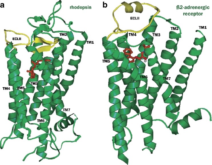
Crystal structures of retinal-bound rhodopsin (a, PDB id 1U19) and carazolol-bound β2-adrenergic receptor (b, PDB id 2RH1). The ligands retinal and carazolol are shown in ball and stick representation with carbon atoms colored brown, while the extracellular loops 2 (ECL2) are shown in yellow. In rhodopsin, ECL2 and the N terminus form a lid over the ligand-binding pocket, whereas in β2-adrenergic receptor the ECL2 contains an extrahelical segment and is more exposed to the solvent, giving open access to the ligand-binding pocket
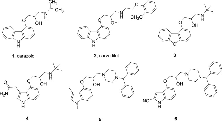
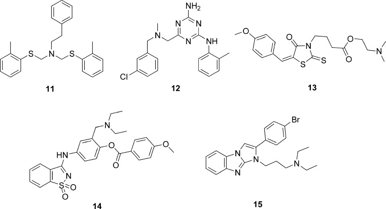
Structures of carazolol, carvedilol, and related ligands predicted to bind like carazolol to β2 adrenergic receptor
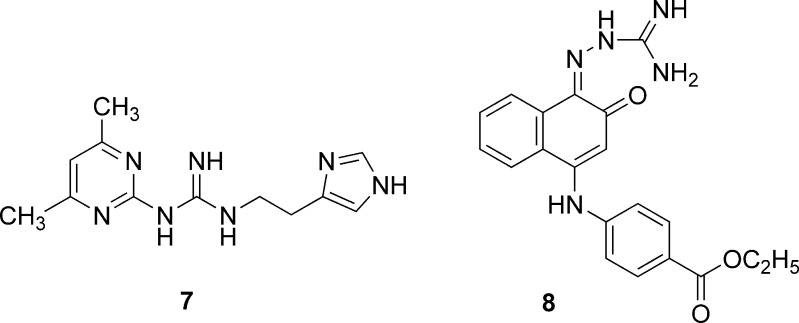
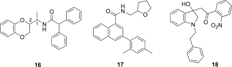
Structures of human histamine H4 receptor identified by virtual screening
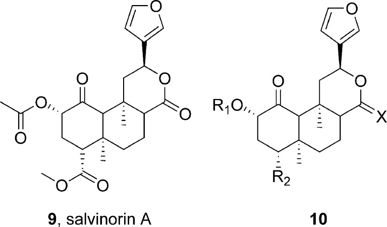
Structures of salvinorin A and related compounds used in pharmacophore modeling and docking at kappa opioid receptor
Structures of antagonist ligands of melanin-concentrating hormone receptor 1 identified by virtual screening
Structures of thyrotropin-releasing hormone receptor 1 antagonists identified by virtual screening
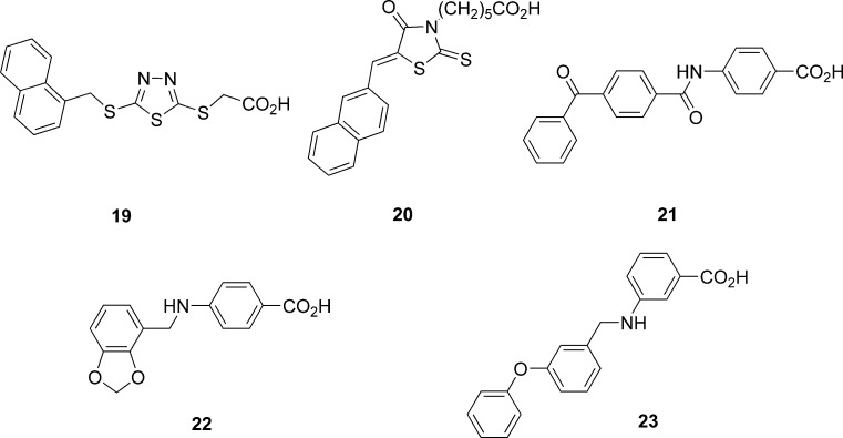
Structures of agonist or partial agonist ligands of the free fatty acid receptor 1 identified by virtual screening
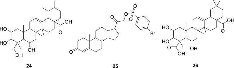
Structures of compounds identified as inhibitors of photoactivated rohodopsin and transducin by virtual screening
Similar articles
-
A benchmarking study on virtual ligand screening against homology models of human GPCRs.Proteins. 2018 Sep;86(9):978-989. doi: 10.1002/prot.25533. Epub 2018 Sep 23. Proteins. 2018. PMID: 30051928
-
Efficiency of Homology Modeling Assisted Molecular Docking in G-protein Coupled Receptors.Curr Top Med Chem. 2021;21(4):269-294. doi: 10.2174/1568026620666200908165250. Curr Top Med Chem. 2021. PMID: 32901584 Review.
-
Modern homology modeling of G-protein coupled receptors: which structural template to use?J Med Chem. 2009 Aug 27;52(16):5207-16. doi: 10.1021/jm9005252. J Med Chem. 2009. PMID: 19627087 Free PMC article.
-
Do crystal structures obviate the need for theoretical models of GPCRs for structure-based virtual screening?Proteins. 2012 Jun;80(6):1503-21. doi: 10.1002/prot.24035. Epub 2012 Mar 13. Proteins. 2012. PMID: 22275072 Free PMC article.
-
X-ray structure breakthroughs in the GPCR transmembrane region.Biochem Pharmacol. 2009 Jul 1;78(1):11-20. doi: 10.1016/j.bcp.2009.02.012. Epub 2009 Feb 27. Biochem Pharmacol. 2009. PMID: 19447219 Review.
Cited by
-
Recent Advances and Applications of Molecular Docking to G Protein-Coupled Receptors.Molecules. 2017 Feb 22;22(2):340. doi: 10.3390/molecules22020340. Molecules. 2017. PMID: 28241450 Free PMC article. Review.
-
Using ligand-based virtual screening to allosterically stabilize the activated state of a GPCR.Chem Biol Drug Des. 2010 Mar;75(3):325-32. doi: 10.1111/j.1747-0285.2009.00944.x. Epub 2010 Jan 19. Chem Biol Drug Des. 2010. PMID: 20659113 Free PMC article.
-
ImmtorLig_DB: repertoire of virtually screened small molecules against immune receptors to bolster host immunity.Sci Rep. 2019 Feb 28;9(1):3092. doi: 10.1038/s41598-018-36179-5. Sci Rep. 2019. PMID: 30816123 Free PMC article.
-
Monte Carlo loop refinement and virtual screening of the thyroid-stimulating hormone receptor transmembrane domain.J Biomol Struct Dyn. 2015;33(5):1140-52. doi: 10.1080/07391102.2014.932310. Epub 2014 Jul 11. J Biomol Struct Dyn. 2015. PMID: 25012978 Free PMC article.
-
How well do the substrates KISS the enzyme? Molecular docking program selection for feruloyl esterases.Sci Rep. 2012;2:323. doi: 10.1038/srep00323. Epub 2012 Mar 20. Sci Rep. 2012. PMID: 22435086 Free PMC article.
References
-
- Cherezov V., Rosenbaum D. M., Hanson M. A., Rasmussen S. G., Thian F. S., Kobilka T. S., Choi H. J., Kuhn P., Weis W. I., Kobilka B. K., Stevens R. C. High-resolution crystal structure of an engineered human beta2-adrenergic G protein-coupled receptor. Science. 2007;318:1258–1265. doi: 10.1126/science.1150577. - DOI - PMC - PubMed
-
- Rosenbaum D. M., Cherezov V., Hanson M. A., Rasmussen S. G., Thian F. S., Kobilka T. S., Choi H. J., Yao X. J., Weis W. I., Stevens R. C., Kobilka B. K. GPCR engineering yields high-resolution structural insights into beta2-adrenergic receptor function. Science. 2007;318:1266–1273. doi: 10.1126/science.1150609. - DOI - PubMed
-
- Rasmussen S. G., Choi H. J., Rosenbaum D. M., Kobilka T. S., Thian F. S., Edwards P. C., Burghammer M., Ratnala V. R., Sanishvili R., Fischetti R. F., Schertler G. F., Weis W. I., Kobilka B. K. Crystal structure of the human beta2 adrenergic G-protein-coupled receptor. Nature. 2007;450:383–387. doi: 10.1038/nature06325. - DOI - PubMed
Publication types
MeSH terms
Substances
LinkOut - more resources
Full Text Sources
