. 2009 Nov;14(6):579-89.
doi: 10.1007/s12192-009-0109-x. Epub 2009 Mar 17.

Heat stress upregulates chaperone heat shock protein 70 and antioxidant manganese superoxide dismutase through reactive oxygen species (ROS), p38MAPK, and Akt

Affiliations

Heat stress upregulates chaperone heat shock protein 70 and antioxidant manganese superoxide dismutase through reactive oxygen species (ROS), p38MAPK, and Akt

Soumyajit Banerjee Mustafi et al. Cell Stress Chaperones. 2009 Nov.

Abstract

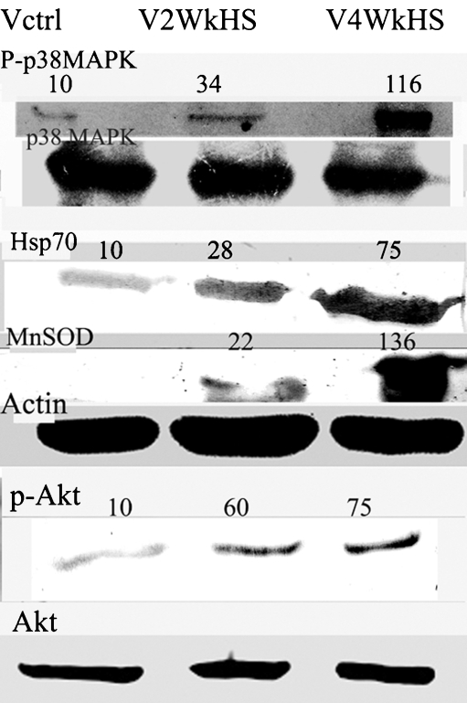
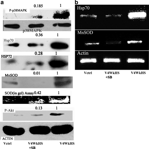
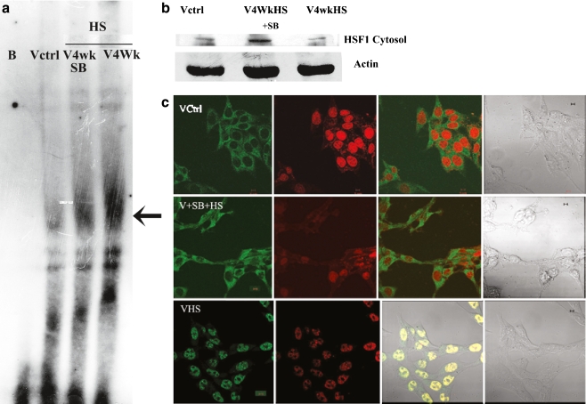
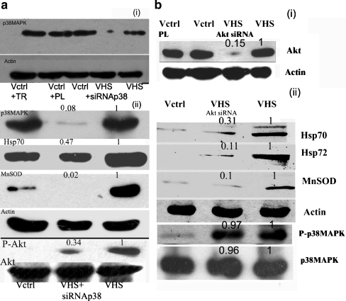
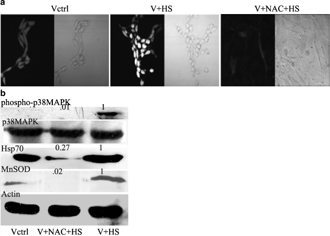
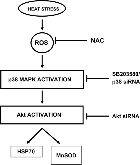
Chinese hamster lung fibroblasts V79 cells were treated with heat stress for 4 weeks with short duration (15 min) heat shock every alternate day in culture. It was observed that Hsp 70 and the antioxidant enzyme MnSOD became overexpressed during the chronic heat stress period. Both p38 MAPK and Akt became phosphorylated by chronic heat stress exposure. Simultaneous exposure to SB203580, a potent and specific p38MAPK inhibitor drastically inhibited the phosphorylation of p38MAPK and Akt. Furthermore, exposure to SB203580 also blocked the increase in Hsp70 and MnSOD levels and the elevated SOD activity brought about by chronic heat stress. Heat shock factor 1 (HSF1) transcriptional activity and nuclear translocation of HSF1 were prominently augmented by chronic heat stress, and this amplification is markedly reduced by concomitant exposure to SB203580. Also, activations of p38MAPK and Akt and upregulations of Hsp70 and MnSOD were observed on exposure to heat shock for a single exposure of longer duration (40 min). siRNA against p38MAPK notably reduced Akt phosphorylation by single exposure to heat stress and drastically diminished the rise in Hsp70 and MnSOD levels. Similarly, siRNA against Akt also eliminated the augmentation in Hsp70 and MnSOD levels but p38MAPK levels remained unaffected. Heat stress produced reactive oxygen species (ROS) in V79 fibroblasts. N-acetyl cysteine blocked the increase in phosphorylation of p38MAPK, amplification of Hsp70, and MnSOD levels by heat stress. Therefore, we conclude that heat stress-activated p38MAPK which in turn activated Akt. Akt acted downstream of p38MAPK to increase Hsp70 and MnSOD levels.Concise summary: Thermal injury of the skin over a long period of time has been associated with development of cancerous lesions. Also, in many cancers, the cytoprotective genes Hsp70 and MnSOD have been found to be overexpressed. Therefore, we considered it important to identify the signaling elements upstream of the upregulated survival genes in heat stress. We conclude that heat stress activated p38MAPK which in turn activated Akt. Akt mediated an augmentation in Hsp70 and MnSOD levels working downstream of p38MAPK.


Figures

Fig. 1
Fig. 1
Chronic heat stress causes increases in phosphorylation of p38MAPK and Akt, and induces enhancement of protein levels of Hsp70 and MnSOD in V79 fibroblasts. Cells were treated with chronic heat stress for a period of 4 weeks. Thermal stress was applied at 43°C for 15 min in culture every alternate day. Cells were harvested after either a 2-week or a 4-week experimental period. Western blotting of the cell lysates was performed. The Western blots are representative of two–three independent experiments. The numbers on the top of the blots are indicative of the respective band densities when the control is normalized to 10. Cells were either left untreated (Vctrl), treated with chronic heat stress (V2WkHS) for 2 weeks, or treated with chronic heat stress (V4WkHS) for 4 weeks. Details of the experimental procedure are given in the materials and methods section
Fig. 2
Fig. 2
Effect of p38MAPK inhibitor SB203580 on the phosphorylation status of p38MAPK and Akt and on the mRNA and protein levels of Hsp70 and MnSOD in V79 fibroblasts subjected to chronic heat stress. a phosphorylation status of p38MAPK (uppermost panel) and Akt (last but one panel from top) and protein expression of Hsp70 (third panel from top), Hsp72 (fourth panel from top), MnSOD (fifth panel from top). SOD enzyme activity is presented in the sixth panel from top. The last panel from top shows beta actin to indicate protein loading. b mRNA level of Hsp70 (uppermost panel), MnSOD (second panel from top), and beta-actin (last panel from top to indicate loading). The numbers on the top of the blots are indicative of the respective band densities when the control is normalized to 1 Details of the experimental procedure are given in the materials and methods section. Cells were either left untreated (Vctrl), treated with chronic heat stress (V4WkHS) or treated simultaneously with chronic heat shock and 5 µM SB203580 (V4WkHS + SB). The results are representative of two-three experiments
Fig. 3
Fig. 3
Effect of SB203580 on the DNA binding activity and cellular localization of heat shock factor 1 (HSF1) in V79 fibroblasts exposed to chronic heat stress. a Electrophoretic Mobility Shift Assay (EMSA) showing HSF1 DNA-binding activity in V79 fibroblasts is provided. Cells were either left untreated (Vctrl), treated with chronic heat stress (V4WkHS), or treated simultaneously with chronic heat shock and 5 µM SB203580 (V4WkHS + SB). Nuclear extracts from these differently treated cells were used for the EMSA. BL Blank without nuclear extract. Arrow indicates HSF1-DNA binding. b Cytosolic localization of HSF1.Upper panel shows HSF1 bands from cytosolic extracts of differently treated cells. Lower panel displays beta-actin to indicate protein loading. Cells were either left untreated (Vctrl), treated with chronic heat stress(V4WkHS) or treated simultaneously with chronic heat shock and 5 µM SB203580 (V4WkHS + SB). c Cellular localization of HSF1 in chronic heat stressed V79 cells by confocal microscopy. Upper panel—Vctrl-untreated V79 cells, middle panel VHS + SB–chronic heat stressed V79 cells treated simultaneously with 5 µM SB203580, lower panel—VHS-chronic heat-stressed cells; micrographs on the left hand side of all panels show cells processed for visualization of HSF1 with secondary antibody–FITC conjugate. The second from left micrographs on all panels show cells processed for visualization of the nucleus with the DNA stain propidium iodide (PI).The third from left micrograph on all panels depict a merger of the FITC and propidium iodide signal. The fourth from left micrograph on all panels show differential interference contrast (DIC) from the same fields. After permeabilization and fixation, cells were treated with anti-HSF1 antibody and subsequently with FITC tagged secondary antibody and PI. The bar is 10 µm. The micrographs are representative of two separate experiments. The cells were treated with chronic heat stress for 4 weeks or were left untreated (control). The chronic heat stressed cells received either no other treatments or received simultaneous treatments with 5 µM SB203580. Experimental details are provided in the “Materials and methods” section
Fig. 4
Fig. 4
Inhibition of the overexpression of Hsp70 and MnSOD induced by heat shock through transfection of siRNA against p38MAPK and Akt. a Exposure to p38MAPK si RNA- (1) Uppermost panel depicts the protein level of p38MAPK. Second panel from top depicts beta actin protein level. (2) Uppermost panel of a series of Western blots depicts the protein level of p38MAPK. Second panel from the top depicts Hsp70. The third panel from the top shows MnSOD levels. The fourth panel from top shows beta actin levels. The fifth panel from top displays phospho-Akt levels. The last panel shows Akt protein levels; Vctrl + TR− control V79 fibroblasts + transfection reagent; VCtrl + PL—Control V79 fibroblasts + Polylinker siRNA; VCtrl- Control V79 fibroblasts; VHS—untransfected V79 fibroblasts treated with single dose heat shock of 43°C for 40 min. VHS + siRNA p38-p38MAPK siRNA transfected V79 fibroblasts treated with single dose heat shock of 43°C for 40 min. b Exposure to Akt siRNA(1) Upper panel shows Akt protein levels, and second panel depicts actin levels. (2) Upper panel depicts Hsp70, the second panel from top depicts Hsp72, and the third panel from the top shows MnSOD immunoblot. The fourth panel from top shows actin immunoblot. The fifth and sixth panels from the top show phospho-p38MAPK and p38MAPK levels, respectively. VCtrl-control V79 fibroblasts; Vctrl + PL-polylinker siRNA treated control; VHS- untransfected V79 fibroblasts treated with single dose heat shock of 43°C for 40 min. VHS + siRNA Akt -siRNA transfected V79 fibroblasts treated with single dose heat shock of 43°C for 40 min. Details of the experimental procedures are described in the “Materials and methods” section. The numbers above the panels indicate band densities with respect to control bands normalized to 1. The blots are representative of two–three experiments
Fig. 5
Fig. 5
Role of ROS on the induction of Hsp70 and MnSOD by heat shock. a Confocal micrographs of V79 fibroblasts treated with single dose heat shock of 43°C for 40 min in the presence and absence of NAC. Cells were grown on coverslips and first incubated with NAC for 2 h. After that, the cell-containing petridishes were given three washes with fresh medium and were subjected to heat shock at 43°C for 40 min. Thereafter, the cells were incubated with the ester form of the H2DCF dye for 15 min at 37°C in Hanks buffer. Cells were visualized under confocal microscope for determining extent of ROS production at the excitation wavelength of 506 nm. Vctrl- untreated cells; V + HS- V79 fibroblasts treated with single dose heat shock of 43°C for 40 min; V + NAC + HS- V79 fibroblasts pretreated with 5 mM NAC and subsequently treated with single dose heat shock of 43°C for 40 min; Vctrl—First picture from left is visualized by dye fluorescence and second picture from the left is differential interference contrast image of the same field. V + HS- Third picture from left is visualized by dye fluorescence and fourth picture from the left is differential interference contrast image of the same field. V + NAC + HS- fifth picture from left is visualized by dye fluorescence and sixth picture from the left is differential interference contrast image of the same field. b Western blot showing p38MAPK phosphorylation and Hsp70 and MnSOD protein levels of V79 fibroblasts treated with single dose heat shock of 43°C for 40 min in the presence and absence of NAC. Vctrl—untreated cells; V + HS- V79 fibroblasts treated with single dose heat shock of 43°C for 40 min; V + NAC + HS- V79 fibroblasts pretreated with 5 mM NAC subsequently treated with single dose heat shock of 43°C for 40 min; The numbers above the panels indicate band densities with respect to control bands normalized to 1. Details of the experimental procedures are described in the “Materials and methods” section
Fig. 6
Fig. 6
Schematic representation of heat stress-induced signaling events leading to the increased expression of Hsp70 and MnSOD. Heat shock induces an enhancement of cellular ROS levels which in turn is responsible for the augmentation of p38MAPK activation. In heat-stressed V79 fibroblasts, Akt is also phosphorylated, and this is controlled by p38MAPK. Activated Akt functions upstream of the overexpression of both Hsp70 and MnSOD

Similar articles

Cited by

References

    1. Arya R, Mallik M, Lakhotia SC. Heat shock genes-integrating cell survival and death. J Biosci. 2007;32:595–610. doi: 10.1007/s12038-007-0059-3. - DOI - PubMed
    1. Aziz SA, Hussain KS, Khan NA, Mushtaq A, Kharadi MY, Bhat JR. Profile of Kangari cancer: a prospective study. Burns. 1998;24:763–766. doi: 10.1016/S0305-4179(98)90094-8. - DOI - PubMed
    1. Calderwood SK, Ciocca DR. Heat Shock Proteins: Stress proteins with Janus-like properties in cancer. Int J Hypertherm. 2008;24:31–39. doi: 10.1080/02656730701858305. - DOI - PubMed
    1. Calderwood SK, Khaleque MA, Sawyer DB, Ciocca DR. Heat shock proteins in cancer:chaperones of tumorigenesis. Trends Biochemic Sci. 2006;31:164–172. doi: 10.1016/j.tibs.2006.01.006. - DOI - PubMed
    1. Chakraborty PK, Banerjee Mustafi S, Ganguly S, Chatterjee M, Raha S. Resveratrol induces apoptosis in K562 (Chronic myelogenous leukemia) cells by targeting a key survival protein Hsp70. Cancer Sc. 2008;99:1109–1116. doi: 10.1111/j.1349-7006.2008.00809.x. - DOI - PMC - PubMed

Publication types

MeSH terms