

High contrast near-infrared polarized reflectance images of demineralization on tooth buccal and occlusal surfaces at lambda = 1310-nm
- PMID: 19291753
- PMCID: PMC2689647
- DOI: 10.1002/lsm.20746
High contrast near-infrared polarized reflectance images of demineralization on tooth buccal and occlusal surfaces at lambda = 1310-nm
Abstract
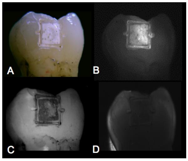
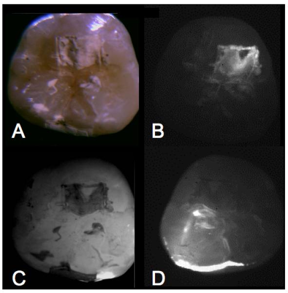
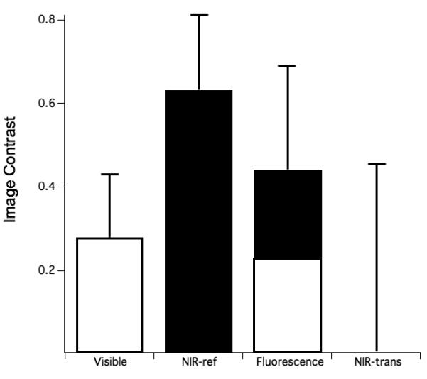
Background and objectives: Sound enamel manifests peak transparency in the near-IR (NIR) at 1310-nm, therefore the near-IR is ideally suited for high contrast imaging of dental caries. The purpose of this study was to acquire images of early demineralized enamel on the buccal and occlusal surfaces of extracted human teeth using NIR reflectance imaging and compare the contrast of those images with the contrast of images taken using other methods.
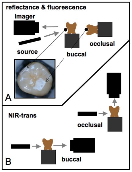
Materials and methods: Fifteen human molars were used in this in vitro study. Teeth were painted with a clear acid-resistant varnish, leaving two 2 mm x 2 mm windows on the buccal and occlusal surfaces of each tooth for demineralization. Artificial lesions were produced in the exposed windows after a 2-day exposure to a demineralizing solution at pH 4.5. Lesions were imaged using NIR transillumination, NIR and visible light reflectance, and fluorescence imaging methods. Crossed polarizers were used where appropriate to improve contrast. Polarization sensitive optical coherence tomography (PS-OCT) was also used to non-destructively assess the depth and severity of demineralization in each sample window.
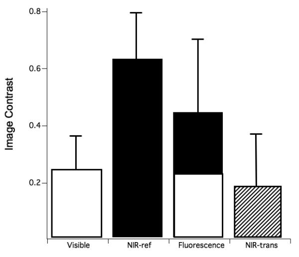
Results: NIR reflectance imaging had the highest image contrast for both the buccal and occlusal groups and it was significantly higher contrast than visible light reflectance (P < 0.05).
Conclusion: The results of the study suggest that NIR reflectance imaging is a promising new method for acquiring high contrast images of early demineralization on tooth surfaces.
Copyright 2009 Wiley-Liss, Inc.
Figures
Similar articles
-
Multispectral near-IR reflectance imaging of simulated early occlusal lesions: variation of lesion contrast with lesion depth and severity.Lasers Surg Med. 2014 Mar;46(3):203-15. doi: 10.1002/lsm.22216. Epub 2013 Dec 27. Lasers Surg Med. 2014. PMID: 24375543 Free PMC article.
-
High contrast reflectance imaging of simulated lesions on tooth occlusal surfaces at near-IR wavelengths.Lasers Surg Med. 2013 Oct;45(8):533-41. doi: 10.1002/lsm.22159. Epub 2013 Jul 16. Lasers Surg Med. 2013. PMID: 23857066 Free PMC article.
-
Imaging Early Demineralization on Tooth Occlusal Surfaces with a High Definition InGaAs Camera.Proc SPIE Int Soc Opt Eng. 2013 Mar 25;8566:85660I-. doi: 10.1117/12.2011015. Proc SPIE Int Soc Opt Eng. 2013. PMID: 24357911 Free PMC article.
-
Early caries imaging and monitoring with near-infrared light.Dent Clin North Am. 2005 Oct;49(4):771-93, vi. doi: 10.1016/j.cden.2005.05.008. Dent Clin North Am. 2005. PMID: 16150316 Review.
-
Transillumination and optical coherence tomography for the detection and diagnosis of enamel caries.Cochrane Database Syst Rev. 2021 Jan 27;1(1):CD013855. doi: 10.1002/14651858.CD013855. Cochrane Database Syst Rev. 2021. PMID: 33502759 Free PMC article.
Cited by
-
Selective removal of demineralized enamel using a CO2 laser coupled with near-IR reflectance imaging.Proc SPIE Int Soc Opt Eng. 2015 Feb 24;9306:93060M. doi: 10.1117/12.2083647. Proc SPIE Int Soc Opt Eng. 2015. PMID: 25914497 Free PMC article.
-
Multispectral near-IR reflectance imaging of simulated early occlusal lesions: variation of lesion contrast with lesion depth and severity.Lasers Surg Med. 2014 Mar;46(3):203-15. doi: 10.1002/lsm.22216. Epub 2013 Dec 27. Lasers Surg Med. 2014. PMID: 24375543 Free PMC article.
-
Automatic method of analysis of OCT images in the assessment of the tooth enamel surface after orthodontic treatment with fixed braces.Biomed Eng Online. 2014 Apr 22;13:48. doi: 10.1186/1475-925X-13-48. Biomed Eng Online. 2014. PMID: 24755213 Free PMC article.
-
Caries Detection Methods Based on Changes in Optical Properties between Healthy and Carious Tissue.Int J Dent. 2010;2010:270729. doi: 10.1155/2010/270729. Epub 2010 Mar 28. Int J Dent. 2010. PMID: 20454579 Free PMC article.
-
Near-Infrared Imaging of Artificial Enamel Caries Lesions with a Scanning Fiber Endoscope.Sensors (Basel). 2019 Mar 22;19(6):1419. doi: 10.3390/s19061419. Sensors (Basel). 2019. PMID: 30909442 Free PMC article.
References
-
-
NIH: Diagnosis and Management of Dental Caries throughout Life 1-24 (NIH Consensus Statement, 2001).
-
-
- Featherstone JDB. Prevention and reversal of dental caries:role of low level fluoride. Community Dent Oral Epidemiol. 1999;27:31–40. - PubMed
-
- Fried D, Featherstone JDB, Glena RE, Seka W. The nature of light scattering in dental enamel and dentin at visible and near-IR wavelengths. Appl. Optics. 1995;34:1278–1285. - PubMed
-
- Jones RS, Fried D. Attenuation of 1310-nm and 1550-nm Laser Light through Sound Dental Enamel, Lasers in Dentistry VIII. Vol. 4610. SPIE; San Jose: 2002. pp. 187–190.
-
- Darling CL, Huynh GD, Fried D. Light Scattering Properties of Natural and Artificially Demineralized Dental Enamel at 1310-nm. J. Biomed. Optics. 2006;11(034023):1–11. - PubMed
Publication types
MeSH terms
Grants and funding
LinkOut - more resources
Full Text Sources
Other Literature Sources
Miscellaneous
