. 2009 Mar 14;15(10):1246-53.
doi: 10.3748/wjg.15.1246.

Morphology and ontogeny of dendritic cells in rats at different development periods

Affiliations

Morphology and ontogeny of dendritic cells in rats at different development periods

Yi-Jun Zhou et al. World J Gastroenterol. .

Abstract

Aim: To study the morphology and ontogeny of dendritic cells of Peyer's patches in rats at different development periods.

Methods: The morphometric and flow cytometric analyses were performed to detect all the parameters of villous-crypts axis and the number of OX62(+)DC, OX62(+)CD4(+)SIRP(+)DC, and OX62(+)CD4(-)SIRP(-)DC in the small intestine in different groups of rats. The relationship between the parameters of villous-axis and the number of DC and DC subtype were analyzed.

Results: All morphometric parameters changed significantly with the development of pups in the different age groups (F = 10.751, 12.374, 16.527, 5.291, 3.486; P = 0.000, 0.000, 0.000, 0.001, 0.015). Villous height levels were unstable and increased from 115.24 microm to 140.43 microm as early as 3 wk postpartum. Villous area increased significantly between 5 and 7 wk postpartum, peeked up to 13817.60 microm(2) at 7 wk postpartum. Villous height and crypt depth ratios were relatively stable and increased significantly from 2.80 +/- 1.01 to 4.54 +/- 1.56, 9-11 wk postpartum. The expression of OX62(+)DC increased from 33.30% +/- 5.80% to 80% +/- 17.30%, 3-11 wk postpartum (F = 5.536, P = 0.0013). OX62(+)CD4(+)SIRP(+)DC subset levels detected in single-cell suspensions of rat total Peyer's patch dendritic cells (PP-DCs) increased significantly from 30.73% +/- 5.16% to 35.50% +/- 4.08%, 5-7 wk postpartum and from 34.20% +/- 1.35% to 43.60% +/- 2.07% 9-11 wk postpartum (F = 7.216, P = 0.005).

Conclusion: This study confirms the age-related changes in villous-crypt axis differentiation in the small intestine. Simultaneously, there are also development and maturation in rat PP-DCs phenotypic expression. Furthermore, the morphological changes of intestinal mucosa and the development of immune cells (especially DC) peaked at 9-11 wk postpartum, indicating that the intestinal mucosae reached a relatively mature state at 11 wk postpartum.


Figures

Figure 1
Figure 1
Comparison of morphological changes of small intestinal mucosa (including villous height, villous width, villous area, crypt depth, and ratio of villous height to crypt depth) at different development periods in SD rats (HE, x 100). A: Group of 3 wk; B: Group of 5 wk; C: Group of 7 wk; D: Group of 9 wk; E: group of 11 wk.
Figure 2
Figure 2
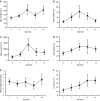
Morphological analysis of villous-crypt axis: All morphological parameters matured as age increased. The results were presented as mean ± SD from 5 rats. A: Villous height increased at 3 wk postpartum, decreased 7 to 9 wk postpartum, and increased again after 9 wk postpartum; B: Villous width increased at 3 wk postpartum, peaked at 7 wk postpartum; C: Villous area increased significantly between 5 and 7 wk postpartum, peaked at 7 wk postpartum; D: Crypt depth increased from 3 to 7 wk postpartum and decreased slightly at 9 wk postpartum; E: Ratio of villous height to crypt depth were relatively stable and increased significantly from 9 to 11 wk postpartum; F: PP increased from 3 to 11 wk postpartum.
Figure 3
Figure 3
FCM of expression of OX62+CD4+SIRP+DC and OX62+CD4-SIRP-DC subsets at different development periods in SD rats. A: Group of 3 wk; B: Group of 5 wk; C: Group of 7 wk; D: Group of 9 wk; E: Group of 11 wk.
Figure 4
Figure 4
Flow cytometric analyses of OX62+CD4+SIRP+ and OX62+CD4-SIRP- PP DCs in vivo at 3, 5, 7, 9 and 11 wk postpartum. The results were presented as mean ± SD from 5 rats. A: Significant growth occurred in different age groups for the number of OX62+DCs; B: Levels of OX62+CD4+OX41+DC subsets increased significantly at 3-5 wk postpartum and 7-9 wk postpartum; C: OX62+CD4-SIRP-DC levels declined on the whole, with small fluctuation from 7 to 9 wk postpartum.

References

    1. Steinman RM, Cohn ZA. Identification of a novel cell type in peripheral lymphoid organs of mice. I. Morphology, quantitation, tissue distribution. J Exp Med. 1973;137:1142–1162. - PMC - PubMed
    1. Steinman RM, Nussenzweig MC. Dendritic cells: features and functions. Immunol Rev. 1980;53:127–147. - PubMed
    1. Spalding DM, Koopman WJ, Eldridge JH, McGhee JR, Steinman RM. Accessory cells in murine Peyer’s patch. I. Identification and enrichment of a functional dendritic cell. J Exp Med. 1983;157:1646–1659. - PMC - PubMed
    1. Spalding DM, Griffin JA. Different pathways of differentiation of pre-B cell lines are induced by dendritic cells and T cells from different lymphoid tissues. Cell. 1986;44:507–515. - PubMed
    1. Iwasaki A, Kelsall BL. Freshly isolated Peyer’s patch, but not spleen, dendritic cells produce interleukin 10 and induce the differentiation of T helper type 2 cells. J Exp Med. 1999;190:229–239. - PMC - PubMed

Publication types