

Human immunodeficiency virus (HIV) antigens and RNA in HIV-seronegative women with cervical intraepithelial neoplasia
- PMID: 19292595
- PMCID: PMC6463994
- DOI: 10.1089/aid.2008.0096
Human immunodeficiency virus (HIV) antigens and RNA in HIV-seronegative women with cervical intraepithelial neoplasia
Abstract
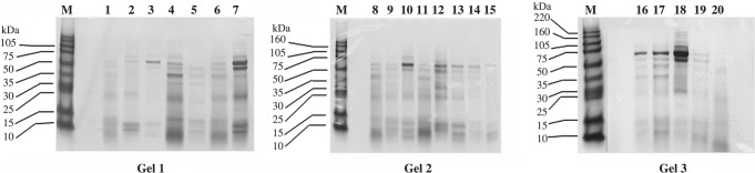
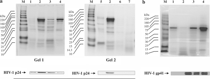
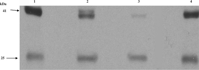
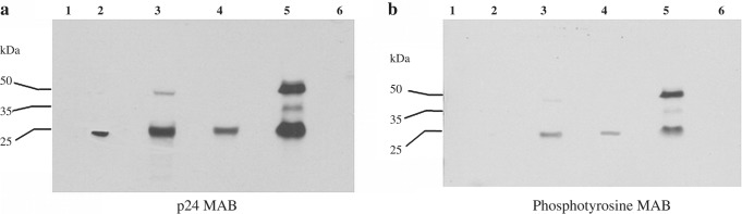
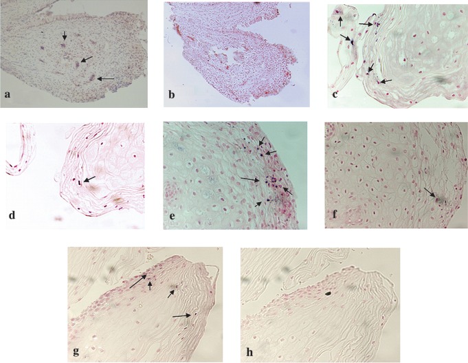
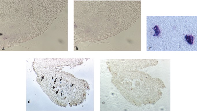
While investigating whether proteins retrieved by cervicovaginal lavages (CVL) from women with cervical intraepithelial neoplasia (CIN) might correlate with risk of progression to invasive cervical cancer, we unexpectedly identified HIV gag and env glycoprotein in CVL from women with HIV-negative serology. HIV antigens were consistently identified by mass spectrometry (MS) in CVL from 4 women but were absent in CVL from the remaining 16 women. HIV serologies of all 20 patients were negative for both HIV-1 and HIV-2 antibodies. To validate the unexpected MS findings we performed Western blot (WB) and immunoaffinity chromatography (IC) analysis of CVL for HIV proteins, viral load assays of paired CVL and blood samples, and immunohistochemical HIV p24 expression in cervical biopsy specimens. WB analysis of CVL for prostate-specific antigen (PSA) was performed to exclude semen contamination as the source of HIV proteins. WB and IC results demonstrated the presence of HIV-1 gp41 and p24 antigens in four CVL that were identified by MS to have the HIV proteins. Despite negative serology, HIV RNA in CVL and HIV p24 in cervix biopsies were detected in patients with HIV antigen-positive CVL. HIV p24-positive CVL were PSA negative. All 20 subjects remained HIV seronegative throughout the study. Women with HIV proteins and RNA were comparatively older. Our findings suggest that CVL HIV proteins in women with CIN could be markers for unrecognized HIV exposure or subclinical infection. Proteomic screening of cervical secretions may be useful in identifying seronegative women exposed to HIV and/or at risk for AIDS.
Conflict of interest statement
No competing financial interests exist.
Figures
Similar articles
-
Scarcity or absence of humoral immune responses in the plasma and cervicovaginal lavage fluids of heavily HIV-1-exposed but persistently seronegative women.AIDS Res Hum Retroviruses. 2011 May;27(5):469-86. doi: 10.1089/aid.2010.0169. Epub 2010 Nov 22. AIDS Res Hum Retroviruses. 2011. PMID: 21091128 Free PMC article.
-
Analysis of HIV-1 in the cervicovaginal secretions and blood of pregnant and nonpregnant women.J Hum Virol. 1999 May-Jun;2(3):154-66. J Hum Virol. 1999. PMID: 10413367
-
Evidence for both Intermittent and Persistent Compartmentalization of HIV-1 in the Female Genital Tract.J Virol. 2019 May 1;93(10):e00311-19. doi: 10.1128/JVI.00311-19. Print 2019 May 15. J Virol. 2019. PMID: 30842323 Free PMC article.
-
Lower genital tract intraepithelial neoplasia in HIV-infected women: guidelines for evaluation and management.Obstet Gynecol Surv. 1999 Feb;54(2):131-7. doi: 10.1097/00006254-199902000-00023. Obstet Gynecol Surv. 1999. PMID: 9950005 Review.
-
Cervical human papillomavirus infection and cervical intraepithelial neoplasia in women positive for human immunodeficiency virus in the era of highly active antiretroviral therapy.Curr Opin Oncol. 2003 Sep;15(5):382-8. doi: 10.1097/00001622-200309000-00007. Curr Opin Oncol. 2003. PMID: 12960521 Review.
Cited by
-
Use of cervicovaginal fluid for the identification of biomarkers for pathologies of the female genital tract.Proteome Sci. 2010 Dec 8;8:63. doi: 10.1186/1477-5956-8-63. Proteome Sci. 2010. PMID: 21143851 Free PMC article.
-
Y chromosome and HIV DNA detection in vaginal swabs as biomarkers of semen and HIV exposure in women.Sex Transm Dis. 2014 Nov;41(11):674-9. doi: 10.1097/OLQ.0000000000000191. Sex Transm Dis. 2014. PMID: 25299415 Free PMC article. Clinical Trial.
References
-
- Broeckel U, Maresso K, and Kugathasan S: Functional genomics and its implications for molecular medicine. Pediatr Clin North Am 2006;53:807–816 - PubMed
-
- Nilsson CL. and Davidsson P: New separation tools for comprehensive studies of protein expression by mass spectrometry. Mass Spectrom Rev 2000;19:390–397 - PubMed
-
- Burk RD, Kadish AS, Calderin S, and Romney SL: Human papillomavirus infection of the cervix detected by cervicovaginal lavage and molecular hybridization: Correlation with biopsy results and Papanicolaou smear. Am J Obstet Gynecol 1986;154:982–989 - PubMed
-
- Basu J, Mikhail MS, Ahn CW, et al. : Plasma uric acid levels in women with cervical intraepithelial neoplasia. Nutr Cancer 2005;51:25–31 - PubMed
-
- Kadish AS, Timmins P, Wang Y, et al. : Regression of cervical intraepithelial neoplasia and loss of human papillomavirus (HPV) infection is associated with cell-mediated immune responses to an HPV type 16 E7 peptide. Cancer Epidemiol Biomarkers Prev 2002;11:483–488 - PubMed
Publication types
MeSH terms
Substances
Grants and funding
LinkOut - more resources
Full Text Sources
Medical
Research Materials
Miscellaneous