. 2009 May;182(1):133-44.
doi: 10.1534/genetics.109.101709. Epub 2009 Mar 16.

A proximal centriole-like structure is present in Drosophila spermatids and can serve as a model to study centriole duplication

Affiliations

A proximal centriole-like structure is present in Drosophila spermatids and can serve as a model to study centriole duplication

Stephanie Blachon et al. Genetics. 2009 May.

Abstract

Most animals have two centrioles in spermatids (the distal and proximal centrioles), but insect spermatids seem to contain only one centriole (Fuller 1993), which functionally resembles the distal centriole. Using fluorescent centriolar markers, we identified a structure near the fly distal centriole that is reminiscent of a proximal centriole (i.e., proximal centriole-like, or PCL). We show that the PCL exhibits several features of daughter centrioles. First, a single PCL forms near the proximal segment of the older centriole. Second, the centriolar proteins SAS-6, Ana1, and Bld10p/Cep135 are in the PCL. Third, PCL formation depends on SAK/PLK4 and SAS-6. Using a genetic screen for PCL defect, we identified a mutation in the gene encoding the conserved centriolar protein POC1, which is part of the daughter centriole initiation site (Kilburn et al. 2007) in Tetrahymena. We conclude that the PCL resembles an early intermediate structure of a forming centriole, which may explain why no typical centriolar structure is observed under electron microscopy. We propose that, during the evolution of insects, the proximal centriole was simplified by eliminating the later steps in centriole assembly. The PCL may provide a unique model to study early steps of centriole formation.


Figures

F<sc>igure</sc> 1.—
Figure 1.—
Ana1 is a centriolar protein required for centriole and cilia formation. (A) Predicted structure of two isoforms (PA and PB) of the Ana1 protein. Red indicates the recognition site of the antibody in the C-terminal part of the protein. The ana1mecB mutation results in a C-to-T transition at position 3358 (relative to ATG), which changes the Q 1120 codon to a stop codon. (B) Centrosomal labeling is reduced in ana1mecB mutants. In the control, GFP–PACT (Martinez-Campos et al. 2004) labels the centriole and γ-tubulin the PCM of mitotic cells from larva brain. In ana1mecB, only 5/25 cells show foci stained by both γ-tubulin and GFP–PACT (arrows), the majority of the cells (18/25) have only γ-tubulin foci, which look smaller than those of the control (double arrows). Few cells (2/25) show a total absence of centrosomal staining. (C) Ana1mecB mutants (grown in 18°) present all the characteristics of ciliary mutants. Flies are uncoordinated, their legs are crossed, and their wings extend upward. EM cross sections of mechanosensory neurons in the control show the tubular body (TB) surrounded by the dendritic sheath (DS). Control images were previously published in Blachon et al. (2008). In ana1mecB, the tubular body is absent but the dentritic sheath is present. At the base of the cilium, basal body (BB) is observed in the control whereas it is absent in ana1mecB. Similarly, in sperm tails no axoneme is detected in ana1mecB whereas in the control they are clearly visible (arrows) sitting near the mitochondria derivatives (arrowheads). (D) In embryos, Ana1-GFP localizes to the centriole surrounded by γ-tubulin. (E) Before meiosis, each spermatocyte contains four giant centrioles arranged by pairs as a V-shape. In primary spermatocytes expressing the centrosomal protein Asl-GFP (Varmark et al. 2007; Blachon et al. 2008), the anti-Ana1 antibody labels the V-shaped giant centrioles. Some nonspecific signal is detected in the nucleus, but in the ana1mecB mutant no giant centriole labeling is detected.
F<sc>igure</sc> 2.—
Figure 2.—
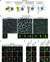
Ana1 labels a novel structure appearing near the mother centriole in spermatids. (A) Diagram depicting the different stages of spermatid development based on the observations of Tates (1971). (M, mitochondria; N, nucleus; Ax, axoneme). The basal body or giant centriole (Cen) is surrounded by the centriolar adjunct (CA) and, near it, we can follow the formation of the PCL. (B) We use phase-contrast pictures (unfixed testis) to determine the spermatid stage. The onion stage (stage S13) is characterized by a round nucleus (N) of the same size as the mitochondrial derivatives (M). The cell body of intermediate spermatids (stages 15 and 16) elongates, forming short protrusions (arrows), but the nucleus remains round. In late spermatid development (stage 17), the nucleus becomes oval. Ana1-GFP labels the giant centriole (Cen), and in intermediate spermatids a bulge forms on one side and becomes individualized as PCL in late spermatid development. (C) Staining with anti-γ-tubulin antibody shows that the PCL labeled by Ana1 is an entity different from the γ-tubulin collar that is reminiscent of the centriolar adjunct (CA). (D) Antibody against Ana1 labels the V-shape pair of giant centrioles in primary spermatocytes (left) and the giant centriole and PCL in spermatids (right) in flies expressing Ana1-GFP. (E) In wild-type primary spermatocytes, anti-Ana1 antibody stains the endogenous protein in the giant centrioles and colocalizes with γ-tubulin staining (left). In spermatids, the antibody labels the PCL, demonstrating that its formation is not due to centriolar protein overexpression (right).
F<sc>igure</sc> 3.—
Figure 3.—
PCL formation depends on early players in centriole duplication. (A and B) PCL formation is impaired in plk4 and sas-6 mutants (for quantification see Table 1). (C) In primary spermatocytes, we can distinguish the daughter centriole (Dau) sitting perpendicularly to the mother centriole (Mo) (based on the γ-tubulin staining). SAS-6-GFP localizes to the proximal part of both mother (Mo) and daughter (Dau) giant centrioles in spermatocytes (left) but is enriched in the daughter. In spermatids, SAS-6-GFP localizes to the proximal side of the giant centriole (Cen) and in addition a second dot, probably in the PCL, appears (right). (D) SAS-6-GFP is localized to the proximal part of the V-shaped pair of giant centrioles in primary spermatocytes (left) stained with anti-Ana1 antibody. In spermatids (right), the staining with anti-Ana1 antibody confirms the colocalization of the SAS-6-GFP second dot with the PCL. (E) We follow the fate of the maternally contributed centriole (for explanation see Blachon et al. 2008) in sas-4 mutants. In sas-4, centrioles are shorter but it does not affect PCL formation, demonstrating that SAS-4 is not required for PCL formation. (F) Like SAS-6-GFP, SAS-4-GFP is localized to the proximal part of the giant centriole in primary spermatocytes. In early spermatids, SAS-4-GFP is still present at the proximal part of the centriole and a second dot can be distinguished. Later, the SAS-4-GFP signal decreased and no SAS4-GFP was detected in the PCL. (G) The PCM protein Cnn shows a similar localization to γ-tubulin (Blachon et al. 2008) in meiosis. However, after meiosis, it is restricted to the proximal part of the giant centriole in early spermatids; subsequently, it is colocalized transiently with the forming PCL in intermediate spermatids. Later, it disappears in late spermatid development. (H) In cnn mutant spermatids, we still observe the PCL labeled by Ana1-GFP, but the γ-tubulin pattern is disrupted. Some spermatids show a very faint or an absence of γ-tubulin labeling (left) whereas others have abnormal localization of the γ-tubulin collars (right).
F<sc>igure</sc> 4.—
Figure 4.—
Bld10p is recruited late to the PCL and is not required for its formation. (A) Bld10p-GFP labels the centriole in spermatocytes (left) and the giant centriole (Cen) in spermatids (right). Only later, in late spermatids, is Bld10p recruited to the PCL. The centriolar adjunct (CA) and the nucleus (N) are visible in phase-contrast pictures (middle). (B) Colocalization of Bld10p-GFP with Ana1-tdTomato confirms that Bld10p localizes to the PCL only later (arrow). The double arrowhead points to the PCL labeled by Ana1-tdTomato and not yet by Bld10p-GFP. To maximize the signal, Bld10p-GFP was expressed in a bld10 mutant context (rescue flies). (C) In the bld10 mutant, centrioles are shorter but PCL formation is not affected, consistent with the role of Bld10p later in the process.
F<sc>igure</sc> 5.—
Figure 5.—
POC1, a conserved centriolar protein, is involved in PCL formation. (A) A forward genetic screen identified a new mutation, k245, that impairs the formation of the PCL as judged by Ana1-GFP labeling. (B) Absence of a normal PCL in k245 is confirmed using Bld10p-GFP. (C) Statistical analysis of the basal body length on poc1 alleles. The deficiency used is Df(3L)Ly. Each experiment was performed on three different flies and 60 basal bodies were measured. The columns marked by a star (*) are statistically different from the control; additional pairs were also found significantly different on the basis of P < 0.01 using Student's t-test. (D) Complementation test with several deletions shows that k245 maps to an interval containing the gene CG10191. Solid bars: deletions that did not complement the k245 phenotype; shaded bars: deletions that did complement k245. (E) CG10191 encodes the conserved centriolar protein POC1. k245 mutation changes a G to an A at position 880 (in red), which is predicted to eliminate a splicing site. poc1c06059 contains an insertion in poc1 promoter (in red). POC1 protein is composed of 7 WD domains known to form a β-propeller structure and contains a coiled-coil domain in its C terminus. The k245 mutation is predicted to induce a frameshift resulting in a truncated protein containing only the WD domains. (F) RT–PCR on mRNA shows that in wild-type flies mRNA is spliced to give a smaller product, which is absent in poc1k245 flies, confirming that the k245 mutation eliminates a splicing site. poc1c06059 shows a significantly lower amount of mRNA than wild type. Actin mRNA is shown as a control for loading.

References

    1. Allen, R. D., 1969. The morphogenesis of basal bodies and accessory structures of the cortex of the ciliated protozoan Tetrahymena pyriformis. J. Cell Biol. 40 716–733. - PMC - PubMed
    1. Andersen, J. S., C. J. Wilkinson, T. Mayor, P. Mortensen, E. A. Nigg et al., 2003. Proteomic characterization of the human centrosome by protein correlation profiling. Nature 426 570–574. - PubMed
    1. Anderson, W. A., 1967. Cytodifferentiation of spermatozoa in Drosophila melanogaster: the effect of elevated temperature on spermiogenesis. Mol. Gen. Genet. 99 257–273. - PubMed
    1. Avidor-Reiss, T., A. M. Maer, E. Koundakjian, A. Polyanovsky, T. Keil et al., 2004. Decoding cilia function: defining specialized genes required for compartmentalized cilia biogenesis. Cell 117 527–539. - PubMed
    1. Azimzadeh, J., and M. Bornens, 2007. Structure and duplication of the centrosome. J. Cell Sci. 120 2139–2142. - PubMed

Publication types

Substances