. 2009 Apr 1;69(7):3077-85.
doi: 10.1158/0008-5472.CAN-08-2281. Epub 2009 Mar 17.

Chemokine expression in melanoma metastases associated with CD8+ T-cell recruitment

Affiliations

Chemokine expression in melanoma metastases associated with CD8+ T-cell recruitment

Helena Harlin et al. Cancer Res. .

Abstract

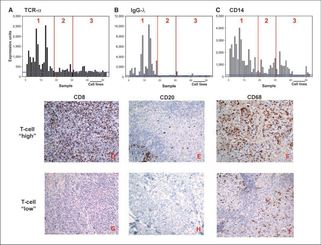
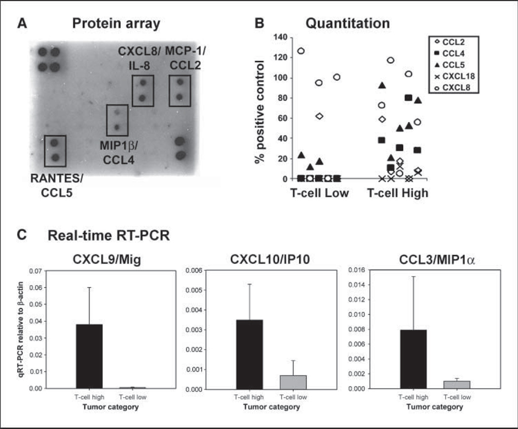
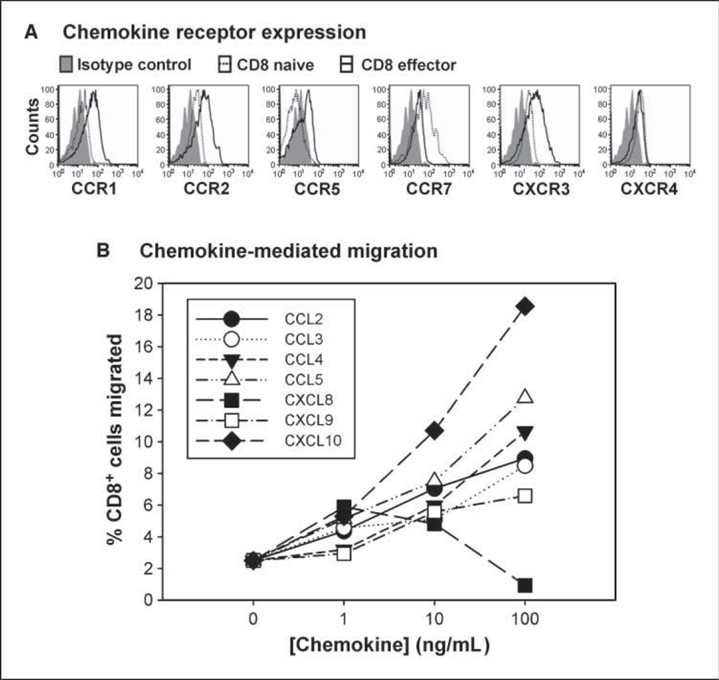
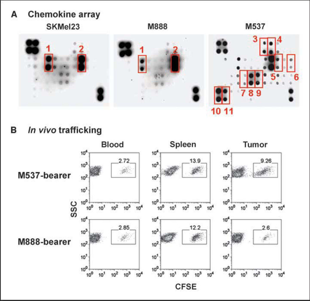
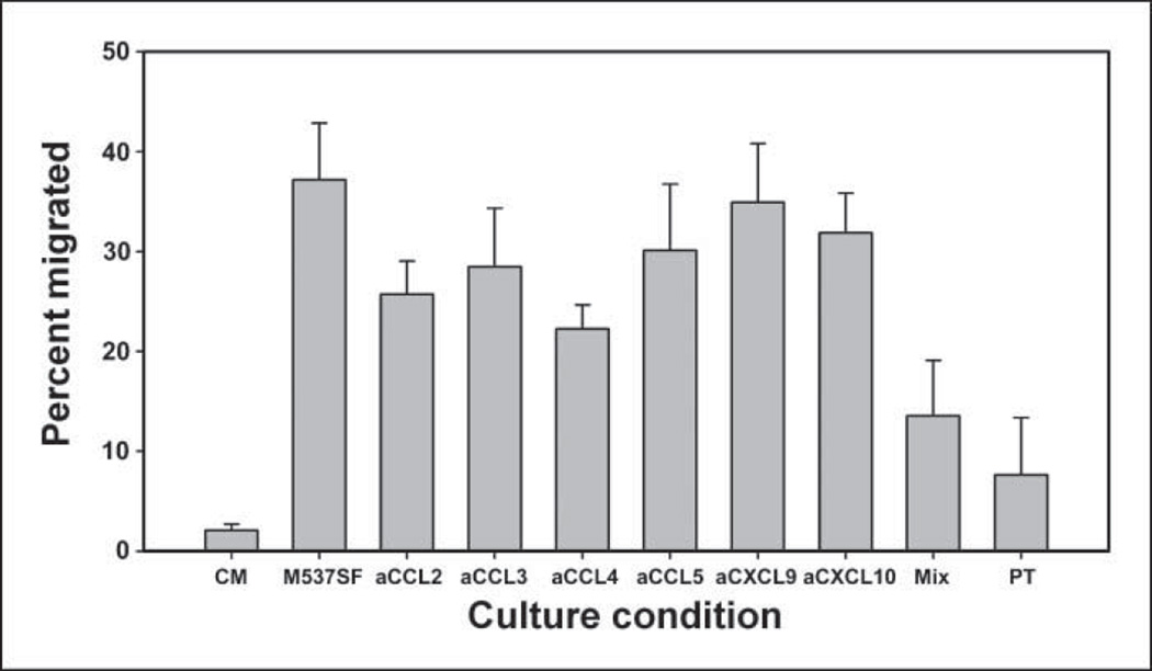
Despite the frequent detection of circulating tumor antigen-specific T cells, either spontaneously or following active immunization or adoptive transfer, immune-mediated cancer regression occurs only in the minority of patients. One theoretical rate-limiting step is whether effector T cells successfully migrate into metastatic tumor sites. Affymetrix gene expression profiling done on a series of metastatic melanoma biopsies revealed a major segregation of samples based on the presence or absence of T-cell-associated transcripts. The presence of lymphocytes correlated with the expression of defined chemokine genes. A subset of six chemokines (CCL2, CCL3, CCL4, CCL5, CXCL9, and CXCL10) was confirmed by protein array and/or quantitative reverse transcription-PCR to be preferentially expressed in tumors that contained T cells. Corresponding chemokine receptors were found to be up-regulated on human CD8(+) effector T cells, and transwell migration assays confirmed the ability of each of these chemokines to promote migration of CD8(+) effector cells in vitro. Screening by chemokine protein array identified a subset of melanoma cell lines that produced a similar broad array of chemokines. These melanoma cells more effectively recruited human CD8(+) effector T cells when implanted as xenografts in nonobese diabetic/severe combined immunodeficient mice in vivo. Chemokine blockade with specific antibodies inhibited migration of CD8(+) T cells. Our results suggest that lack of critical chemokines in a subset of melanoma metastases may limit the migration of activated T cells, which in turn could limit the effectiveness of antitumor immunity.


Conflict of interest statement

Disclosure of Potential Conflicts of Interest

No potential conflicts of interest were disclosed.

Figures

Figure 1
Figure 1
Distribution of selected inflammatory cells’ transcripts in melanoma biopsies. A to C, gene array data. Expression levels of TCRα (indicating T-lineage cells), IgGλ (indicating B-lineage cells), and CD14 (indicating monocyte-lineage cells) transcripts represented as normalized hybridization intensity data are shown for individual samples. The vertical lines indicate separations between groups 1, 2, and 3, and the horizontal line indicates the data with melanoma cell lines. D to I, immunohistochemical confirmation of inflammatory cell infiltrates. Representative tissue samples from tumors that contained (top) or lacked (bottom) T-cell transcripts were stained with antibodies specific for CD8 (D and G), CD20 (E and H), and CD68 (F and I). Similar results were observed with two additional tumors from each group.
Figure 2
Figure 2
Correlation between chemokines and T-cell transcripts in individual tumors. A, expression levels of CCL5 transcripts represented as normalized hybridization intensity data are shown for individual samples. The vertical lines indicate separations between groups 1, 2, and 3. B, plot of CCL5 transcript levels versus TCRα levels in individual tumors. A positive correlation was observed (R2 = 0.6). C, nonsupervised hierarchical clustering of chemokine and CD8β transcript data. To examine associations between CD8 expression and a diverse panel of chemokines, nonsupervised hierarchical clustering analysis was done on the subset of transcripts that encode chemokines and CD8β. CD8β was found to spontaneously cluster with a subset of chemokine transcripts (indicated by the box).
Figure 3
Figure 3
Quantitative expression of chemokine proteins in melanoma metastases that contain or lack T cells. Antibody-based protein arrays were used to assess the presence of 38 chemokines in tumor lysates. A, representative blots from a tumor rich in T-cell transcripts versus a tumor that lacked T-cell transcripts. B, quantitative data from scanned blots from five tumors that contained T-cell transcripts and five tumors that lacked T-cell transcripts. C, real-time RT-PCR was done for CXCL9, CXCL10, and CCL3 on five tumors that contained or lacked T-cell transcripts. Columns, mean; bars, SD.
Figure 4
Figure 4
Chemokines relevant for recruitment of CD8+ effector T cells. A, chemokine receptor expression. Flow cytometric analysis was done comparing naive versus effector CD8+ T cells obtained from normal donors for expression of CCR1, CCR2, CCR5, CXCR3, CCR7, and CXCR4. Similar results were seen with at least two independent donors, and with effector cells generated by in vitro priming with anti-CD3/anti-CD28 mAb–coated beads. B, chemokine-mediated migration. In vitro transwell migration was done using in vitro primed CD8+ effector cells in the upper chamber and the indicated chemokines in the lower chamber. The percentages of T cells in the lower chamber were determined by flow cytometry at 2 h and are representative of at least two experiments.
Figure 5
Figure 5
Chemokines produced by a subset of melanoma cell lines can attract human CD8+ effector T cells in a xenograft setting in vivo. A, supernatants from a series of melanoma cell lines were examined for chemokine content using a protein array. Melanoma cell line M537 showed a more diverse chemokine production profile. The indicated chemokines are GRO (1), IL-8 (2), CTACK (3), CXCL16 (4), IP-10 (5), MCP-1 (6), MIG (7), MIP-1α (8), MIP-1β (9), PARC (10), and RANTES (11). Each sample is represented in duplicate. The four top left corner spots and two bottom right corner spots represent loading controls. B, human melanoma cell lines were implanted s.c. into NOD/scid mice. Once they grew to a solid tumor, CD8+ effector T cells were prepared from normal human donors, labeled with CFSE as a fluorescent indicator, and injected i.v. The indicated tissues were then harvested and analyzed by flow cytometry for the presence of labeled T cells versus side scatter. Similar results were seen in at least two independent experiments.
Figure 6
Figure 6
Chemokine blockade inhibits recruitment of CD8+ effector T cells by M537 tumor-derived supernatants. Supernatants were generated from M537 tumor cells and assessed for the ability to attract human CD8+ effector T cells from normal donors. Migration was analyzed in the presence of the indicated chemokine-specific antibodies or with pertussis toxin pretreatment of the T cells (PT). Culture medium alone (CM) was used as a negative control. Similar results were seen in at least two experiments.

References

    1. Rosenberg SA. Cancer vaccines based on the identification of genes encoding cancer regression antigens. Immunol Today. 1997;18:175–182. - PubMed
    1. Van den Eynde B, Gaugler B, van der Bruggen P, Coulie P, Brichard V, Boon T. Human tumour antigens recognized by T-cells: perspectives for new cancer vaccines. Biochem Soc Trans. 1995;23:681–686. - PubMed
    1. Peterson AC, Harlin H, Gajewski TF. Immunization with Melan-A peptide-pulsed peripheral blood mononuclear cells plus recombinant human interleukin-12 induces clinical activity and T-cell responses in advanced melanoma. J Clin Oncol. 2003;21:2342–2348. - PubMed
    1. Cormier JN, Salgaller ML, Prevette T, et al. Enhancement of cellular immunity in melanoma patients immunized with a peptide from MART-1/Melan A. Cancer J Sci Am. 1997;3:37–44. - PMC - PubMed
    1. Rosenberg SA, Sherry RM, Morton KE, et al. Tumor progression can occur despite the induction of very high levels of self/tumor antigen-specific CD8+ T cells in patients with melanoma. J Immunol. 2005;175:6169–6176. - PubMed

Publication types