. 2009 Apr 1;69(7):2845-52.
doi: 10.1158/0008-5472.CAN-08-2089. Epub 2009 Mar 17.

The membrane mucin Muc4 inhibits apoptosis induced by multiple insults via ErbB2-dependent and ErbB2-independent mechanisms

Affiliations

The membrane mucin Muc4 inhibits apoptosis induced by multiple insults via ErbB2-dependent and ErbB2-independent mechanisms

Heather C Workman et al. Cancer Res. .

Abstract

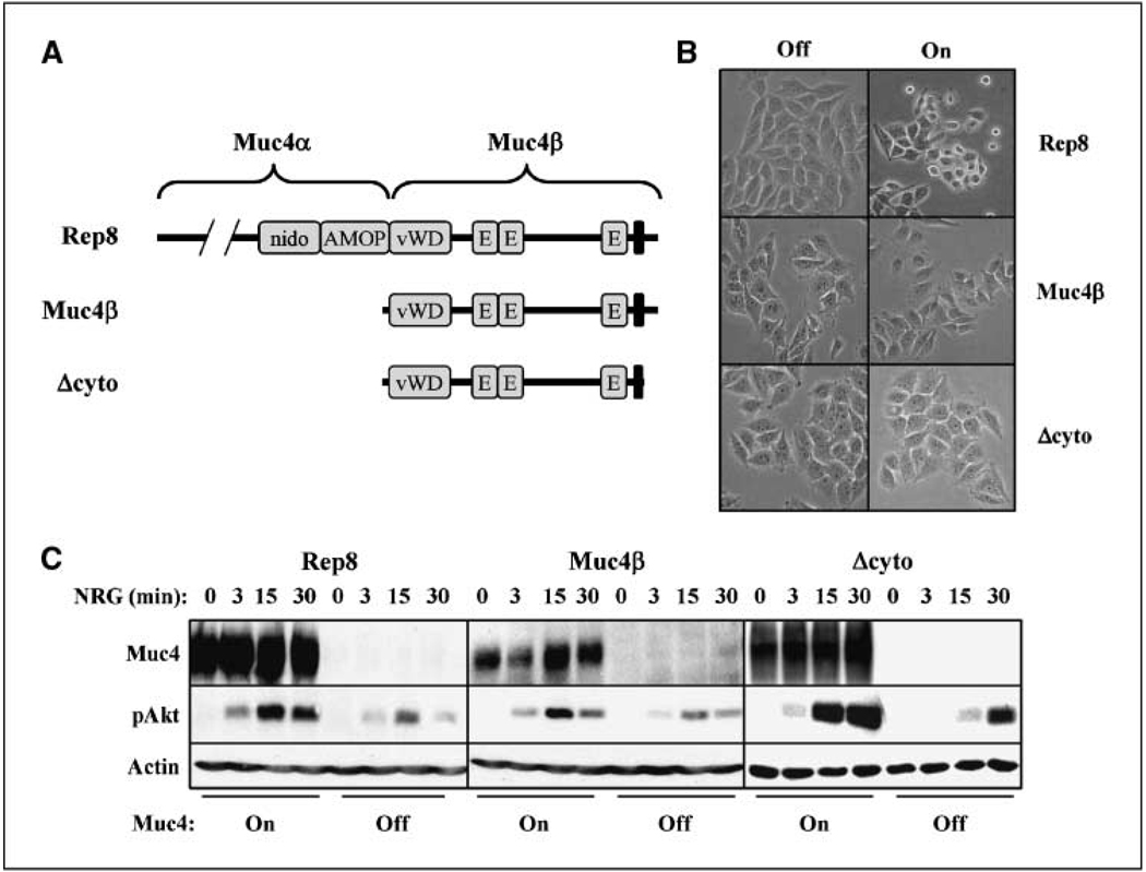
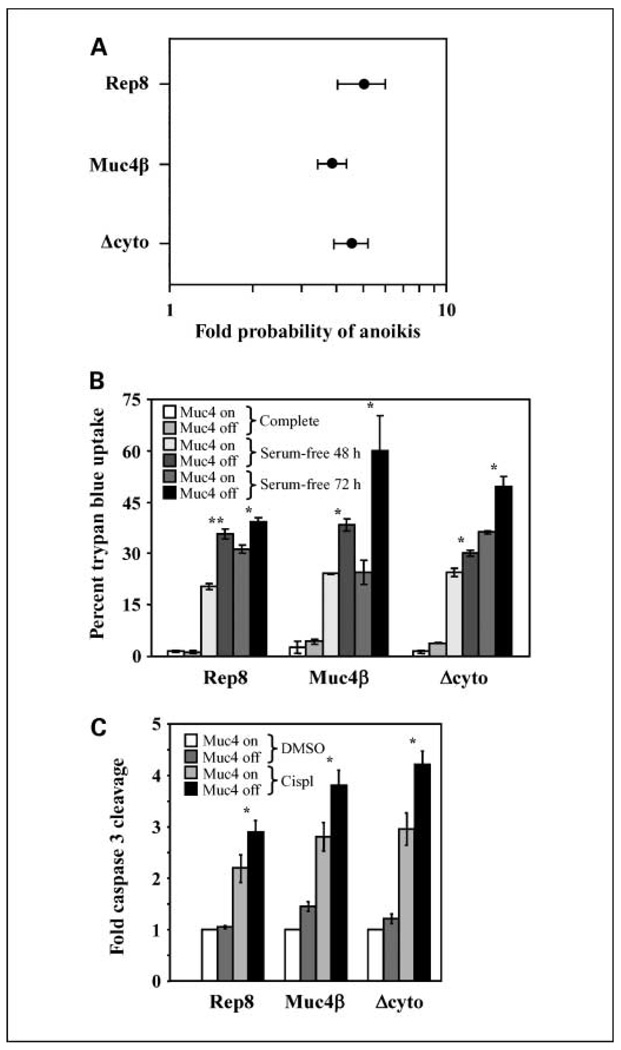
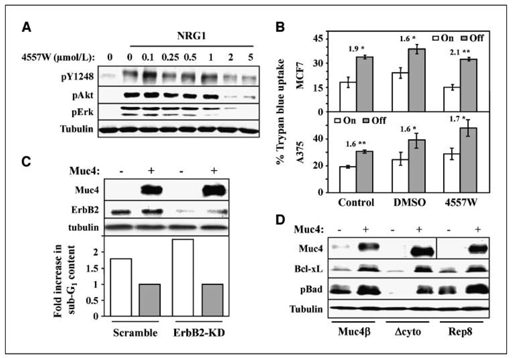
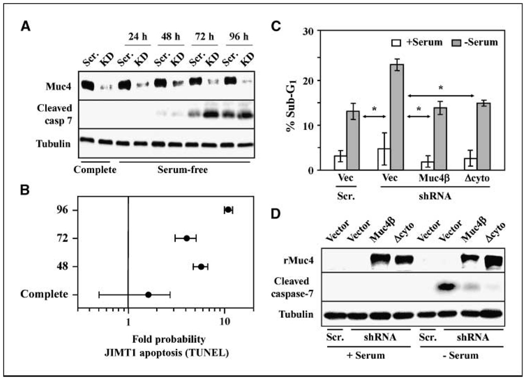
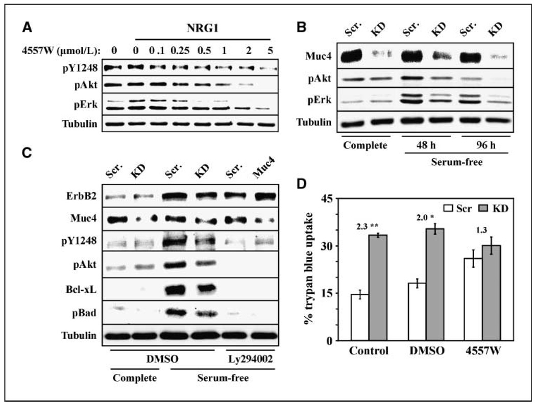
The aberrant expression of membrane mucins such as Muc1 and Muc4 by tumor cells has been shown to engage signaling pathways that promote cellular properties associated with tumor progression. Our previous studies have shown that Muc4 interacts with and potentiates signaling by the ErbB2 (HER2) receptor tyrosine kinase through an epidermal growth factor-like domain in its extracellular region. Here, we show that expression of Muc4 in human A375 melanoma cells and MCF7 breast cancer cells confers resistance to apoptosis induced by a variety of stimuli, including chemotherapeutic agents, the absence of serum factors, and the loss of cellular adhesion. Mapping experiments revealed that the O-glycosylation and cytosolic domains of Muc4 are dispensable for its antiapoptotic activity, and are also dispensable for the potentiation of signaling by ErbB2. Knockdown of endogenous Muc4 in JIMT-1 breast cancer cells sensitizes cells to apoptotic stimuli, and this can be rescued by Muc4 forms lacking the O-glycosylation or cytosolic domains. Surprisingly, however, the molecular mechanisms underlying Muc4 antiapoptotic activity vary among cell lines. Although Muc4 in JIMT-1 cells engages ErbB2 to promote cell survival, its antiapoptotic mechanism in MCF7 and A375 cells seems to be independent of ErbB2. However, Muc4 expression in all cell lines culminates in the phosphorylation and inactivation of the proapoptotic protein Bad and the elevation of the prosurvival protein Bcl-xL. Our observations suggest that tumor cells can exploit the versatile antiapoptotic activities of Muc4 to acquire resistance to therapeutic agents, and augment cell survival after the loss of adhesion and microenvironment-derived survival factors.


Conflict of interest statement

Disclosure of Potential Conflicts of Interest

No potential conflicts of interest were disclosed.

Figures

Figure 1
Figure 1
The cytosolic and O-glycosylation domains are dispensable for Muc4-potentiated signaling by ErbB receptors. A, the domain structures of the Muc4 forms used are depicted. The illustrated domains in rat Rep8 are as follows: amino terminal O-glycosylation, nidogen, AMOP, von Willebrand D, three EGF-like domains, a transmembrane segment, and a short cytoplasmic tail. B, A375 cells inducibly expressing rat full-length Muc4 (Rep8),Muc4β, or Δcyto were treated with and without tetracycline for 72 h to induce expression, and were photographed. C, cell lines were treated without and with tetracycline for 48 h, and then treated with NRG1 for the indicated times. Cell lysates were blotted with antibodies to Muc4, phospho-Akt, and actin.
Figure 2
Figure 2
The cytosolic and O-glycosylation domains are dispensable for Muc4 antiapoptotic activity. A, A375 cells inducibly expressing Rep8, Muc4β, or Δcyto were suspended for 72 h, and anoikis was measured by TUNEL followed by FACS. The fold increase in the probability that the cells will undergo apoptosis in the absence of Muc4 relative to Muc4 expression was calculated and plotted for each of the three constructs. In the Forest meta-analysis plot, 1 represents no effect and 10 represents a 10-fold higher probability as calculated by odds ratio, with a P value of <0.001. Error bars, the 95% confidence interval for the population; points, the center of the confidence interval. B, inducible cell lines were grown in serum-free media for 48 or 72 h, as indicated, and apoptosis was measured by trypan blue uptake. C, cells with Muc4 forms turned on or off were treated with DMSO vehicle or with 5.5 µg/mL cisplatin (cispl ) for 18 h, cleaved caspase 3 levels were determined by immunoblotting and bands were quantified digitally. The fold enhancement of cleaved caspase levels over levels observed in the presence of Muc4 but the absence of drug is plotted for each cell line. *, P < 0.05; **, P < 0.001.
Figure 3
Figure 3
ErbB2 is dispensable for antiapoptotic activity in MCF7 and A375 cells. A, MCF7-Rep5 cells were serum starved overnight and treated without and with NRG1 for 10 min, as indicated, and with increasing concentrations of 4557W. Cell lysates were immunoblotted with the indicated antibodies. B, MCF7-Rep5 cells (top) or A375-Rep8 cells growing in serum-free media for 48 h were left untreated or were treated with DMSO control or 2 µmol/L 4557W, as indicated, in the presence or absence of Muc4 expression. The percentage of viable cells was determined by trypan blue exclusion. The fold increased apoptosis in the absence relative to the presence of Muc4 expression is indicated. *, P < 0.05; **, P < 0.001. C, MCF7-Rep5 cells were transfected with scrambled or ErbB2-directed knockdown oligonucleotides, and were simultaneously treated with and without tetracycline for 48 h to induce Muc4 expression. Lysates were blotted with the indicated antibodies (top). Apoptosis for the indicated conditions was determined by sub-G1 analysis, and the fold increase in apoptosis in the absence of Muc4 relative to its presence is plotted (bottom). D, lysates from A375-Rep8, Muc4β, and Δcyto cells grown in the absence of serum for 72 h were blotted with antibodies to Muc4, Bcl-xL, phosphor-Bad, and tubulin.
Figure 4
Figure 4
Endogenous Muc4 promotes the viability of JIMT-1 breast tumor cells. A, JIMT-1 cells stably transduced with retrovirus expressing a Muc4-directed shRNA construct (KD) or a scrambled control (Scr.) were grown in complete or serum-free media for the indicated times. Lysates were immunoblotted with antibodies to Muc4, cleaved caspase 7 (cleaved casp 7),and tubulin. B, JIMT-1 cells transduced with scrambled or Muc4-directed shRNA constructs were grown in serum-free media for the indicated times, and the extent of apoptosis was determined by TUNEL followed by FACS. The fold increase in the probability that the cells will undergo apoptosis when Muc4 is knocked down relative to Muc4 expression was calculated and plotted for three time points. C, wild-type JIMT-1 cells were transiently cotransfected with either Muc4-directed shRNA or scrambled vectors along with Muc4β, Δcyto, or control (vec) plasmids, as indicated. Cells grown in the presence and absence of serum were assayed for apoptosis by sub-G1 content. *, P < 0.05 relative to shRNA+vector with no serum. D, whole cell lysates from cells depicted in C were blotted with the indicated antibodies.
Figure 5
Figure 5
Muc4-induced survival is mediated by ErbB2 signaling in JIMT-1 cells. A, JIMT-1-Scr cells grown in serum-free media overnight were treated without and with NRG1 for 10 min, as indicated, and with increasing concentrations (0–5 µmol/L) of 4557W ErbB inhibitor. Lysates were blotted with phospho-ErbB2 (pY1248),phospho-Akt, phospho-Erk, and tubulin antibodies. B, lysates from JIMT-1-Scr and JIMT-1-KD cells grown in complete or serum-free media for the indicated times were blotted with the indicated antibodies. C, JIMT-1-Scr and JIMT-1-KD cells grown in complete or serum-free media for 72 h were treated with DMSO control or 20 µmol/L Ly294002. Lysates were blotted with the indicated antibodies. D, JIMT-1-Scr and JIMT-1-KD cells grown in serum-free media for 72 h were left untreated or treated with DMSO or 4 µmol/L 4557W ErbB inhibitor, as indicated, and the percentage cells exhibiting trypan blue uptake was determined. The fold increase in apoptosis in the absence relative to the presence of Muc4 expression is indicated. *, P < 0.05; **, P < 0.001.

References

    1. Chaturvedi P, Singh AP, Batra SK. Structure, evolution, and biology of the MUC4 mucin. FASEB J. 2008;22:966–981. - PMC - PubMed
    1. Hattrup CL, Gendler SJ. Structure and function of the cell surface (tethered) mucins. Annu Rev Physiol. 2008;70:431–457. - PubMed
    1. Carraway KL, III, Funes M, Workman HC, Sweeney C. Contribution of membrane mucins to tumor progression through modulation of cellular growth signaling pathways. Curr Top Dev Biol. 2007;78:1–22. - PubMed
    1. Cullen PJ. Signaling mucins: the new kids on the MAPK block. Crit Rev Eukaryot Gene Expr. 2007;17:241–257. - PubMed
    1. Singh PK, Hollingsworth MA. Cell surface-associated mucins in signal transduction. Trends Cell Biol. 2006;16:467–476. - PubMed

Publication types