Multicenter Study
. 2009 Jun;132(Pt 6):1601-12.
doi: 10.1093/brain/awp049. Epub 2009 Mar 17.

Distinct phenotypic and functional features of CADASIL mutations in the Notch3 ligand binding domain

Affiliations
Multicenter Study

Distinct phenotypic and functional features of CADASIL mutations in the Notch3 ligand binding domain

Marie Monet-Leprêtre et al. Brain. 2009 Jun.

Abstract

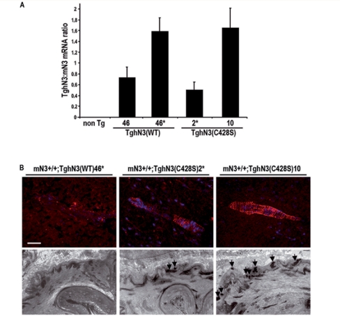
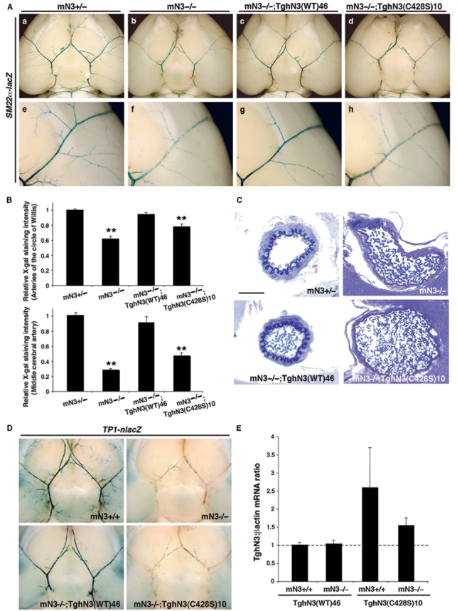
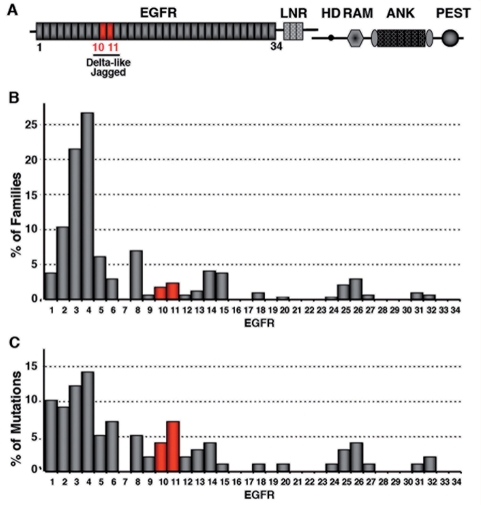
Cerebral autosomal dominant arteriopathy with subcortical infarcts and leukoencephalopathy (CADASIL) is an autosomal dominant small-vessel disease of the brain caused by mutations in the NOTCH3 receptor. The highly stereotyped nature of the mutations, which alter the number of cysteine residues within the epidermal growth factor-like repeats (EGFR), predicts that all mutations share common mechanisms. Prior in vitro assays and genetic studies in the mouse support the hypothesis that common mutations do not compromise canonical Notch3 function but instead convey a non-physiological and deleterious activity to the receptor through the unpaired cysteine residue. Intriguingly, in vitro studies predict that mutations located in the Delta/Serrate/LAG-2 ligand binding domain-(EGFR10-11) may result in a loss of Notch3 receptor function. However, the in vivo relevance and functional significance of this with respect to the pathogenic mechanisms and clinical expression of the disease remain largely unexplored. To ascertain, in vivo, the functional significance of EGFR10-11 mutations, we generated transgenic mice with one representative mutation (C428S) in EGFR10 of Notch3. These mice, like those with a common R90C mutation, developed characteristic arterial accumulation of Notch3 protein and granular osmiophilic material upon aging. By introducing the mutant C428S transgene into a Notch3 null background, we found that, unlike the R90C mutant protein, the C428S mutant protein has lost wild-type Notch3 activity and exhibited mild dominant-negative activity in three different biological settings. From a large prospectively recruited cohort of 176 CADASIL patients, we identified 10 patients, from five distinct pedigrees carrying a mutation in EGFR10 or 11. These mutations were associated with significantly higher Mini-Mental State Examination and Mattis Dementia Rating Scale scores (P < 0.05), when compared with common mutations. Additionally, we found a strong effect of this genotype on the burden of white matter hyperintensities (P < 0.01). Collectively, these results highlight distinctive functional and phenotypic features of EGFR10-11 mutations relative to the common CADASIL mutations. Our findings are compatible with the hypothesis that EGFR10-11 mutations cause the disease through the same gain of novel function as the common mutations, and lead us to propose that reduced Notch3 signalling acts as a modifier of the CADASIL phenotype.


Figures

Figure 1
Figure 1
Generation and characterization of TghN3(C428S) mice. (A) Expression level of human NOTCH3 transgenes in isolated brain arteries from wild-type [TghN3(WT)] and mutant [TghN3(C428S)] transgenic mice on a normal Notch3+/+ background, at 1 month of age. Total RNA was prepared from brain arteries collected from 2 to 3 mice. Lines 46 and 46* carry the wild-type transgene at the heterozygous and homozygous state respectively. Lines 2* and 10 are independent lines carrying the mutant transgene at the heterozygous and homozygous state respectively. Shown are mean ratios (±SEM) of human NOTCH3 mRNA to murine Notch3 mRNA measured in 3–4 RNA preparations for each line as determined by real-time RT–PCR. (B) Upper panel, shown are representative brain artery sections from 10-month-old TghN3(WT)46*, 18-month-old TghN3(C428S)2* and 8-month-old TghN3(C428S)10 mice on a Notch 3+/+ background, immunostained with the 1E4 mAb against human NOTCH3ECD (dapi nuclear staining). TghN3(C428S) mice from both lines exhibit the characteristic granular Notch3 immunostaining, indicative of accumulation of aggregated NOTCH3ECD whereas TghN3(WT) mice show no staining in the same experimental conditions. Lower panel, representative electron micrographs of brain arteries from 10-month-old TghN3(WT)46*, 18-month-old TghN3(C428S)2* and 8-month-old TghN3(C428S)10 mice on a Notch 3+/+ background showing granular osmiophilic material deposits (arrows) in TghN3(C428S) arteries whereas granular osmiophilic material deposits are absent in TghN3(WT) artery. Scale bar: 35 µm (upper panel), 0.7 µm (lower panel).
Figure 2
Figure 2
The C428S mutation creates a non-functional allele. (A) One-month-old mice of the indicated line and genotype heterozygous for the SM22α-lacZ transgene, were assayed for smooth muscle cell arterial identity with X-gal staining on whole brain. Shown are ventral views of whole brain with the circle of Willis and its main branches (a–d) and higher magnification views of the middle cerebral artery (e–h). mN3−/−,TghN3(WT) rescued mice have comparable X-gal staining in the large and middle size brain arteries (c and g) to that seen in the Notch3+/− control mice (a and e). In contrast, X-gal staining in mN3−/−, TghN3(C428S) mice is mildly reduced in the arteries of the circle of Willis (d) and strongly reduced in the middle cerebral artery (h) as seen in Notch3−/− mice (b and f). (B) Relative SM22α-lacZ transgene expression level in the arteries of the circle of Willis (upper panel) and the middle cerebral artery (MCA) (lower panel) of 1-month-old mice heterozygous for the SM22α-lacZ transgene of the following genotypes: Notch3+/- (n = 15 mice), Notch3−/- (n = 10 mice), mN3−/−;TghN3(WT)46 (n = 5 mice) and mN3−/-;TghN3(C428S)10 (n = 7 mice) in the upper panel and Notch3+/− (n = 25 MCA from 13 mice), Notch3−/− (n = 20 MCA from 12 mice), mN3−/−;TghN3(WT)46 (n = 9 MCA from 5 mice), and mN3−/−;TghN3(C428S)10 (n = 10 MCA from five mice) in the lower panel. Shown are mean intensities of X-gal staining (±SEM) in arteries expressed relative to those in Notch3+/- mice arbitrarily set to a value of 1. Differences were evaluated by ANOVA and Scheffe's test (**P < 0.001 versus Notch3+/- or mN3−/−;TghN3(WT)46 mice). (C) Shown are representative semi-thin sections of brain arteries stained with toluidine blue from mice of the indicated line and genotype, aged one month. Brain artery in mN3−/-; TghN3(C428S) mice is enlarged, with a flattened elastica lamina and abnormally thin smooth muscle cells as seen in Notch3−/− mice. Scale bar: 25 µm. (D) One-month-old mice of the indicated genotype, carrying the TP1-nlacZ transgene at the heterozygous state, were assayed for RBP-Jκ activity, with X-gal staining on dissected whole-brain. Shown are ventral views of whole brains. Robust X-gal staining is detected in arteries of the Notch3+/+ and mN3−/−;TghN3(WT) rescued mice while vascular staining is similarly abrogated in mN3−/−;TghN3(C428S) and Notch3−/− mice. (E) Relative expression levels of human NOTCH3 transgenes in isolated brain arteries (circle of Willis and its main branches) from wild-type TghN3(WT)46 and mutant TghN3(C428S)10 transgenic mice on Notch3+/+ and Notch3−/− backgrounds, at one month of age. Expression level of hNOTCH3 transgenes was normalized for β-actin and expressed relative to the level in mN3+/+;TghN3(WT) arteries and arbitrarily set to a value of 1.
Figure 3
Figure 3
Dominant negative effect of the C428S mutant NOTCH3 protein. One-month-old mN3+/−;TghN3(C428S)10 and control mice were assayed for smooth muscle cell arterial identity (A), brain arterial morphology and smooth muscle cell ultrastructure (B) and Notch3/RBP-Jκ activity (C). (A) Left panel: lateral views of hemisphere from mice carrying the SM22α-lacZ transgene, showing reduced X-gal staining in the middle cerebral artery and its branches in TghN3(C428S) mice on a Notch3+/− background as compared to control Notch3+/− mice. Right panel: Relative β-galactosidase activity in the middle cerebral artery of mN3+/−;TghN3(C428S)10 (n = 6 arteries from three mice) and Notch3+/− mice (n = 25 arteries from 13 mice) heterozygous for the SM22α-lacZ transgene. Shown are mean intensities of blue X-gal staining (±SEM) expressed relative to those in Notch3+/− mice arbitrarily set to a value of 1. (*P < 0.001 versus Notch3+/− mice; unpaired Student's t-test). (B) Shown are representative semi-thin sections of brain arteries stained with toluidine blue (a and b) and representative electron micrographs (c and d) showing focal alterations of the arterial vessel with flattened elastica lamina (e.l.), smooth muscle cell (SMC) thinning and vacuolization (arrowheads) in mN3+/−; TghN3(C428S). Scale bar: 20 µm (a and b), 2.9 µm (c and d). (C) Shown are ventral views of whole brain from mice of the indicated genotype carrying the TP1-nlacZ transgene at the heterozygous state. X-gal staining of brain arteries in TghN3(C428S) mice on a Notch3+/− background is significantly reduced as compared to Notch3+/− control mice and mildly reduced in TghN3(C428S) mice on a Notch3+/+ background as compared to Notch3+/+ control mice.
Figure 4
Figure 4
Distribution of CADASIL-associated NOTCH3 mutations across EGFR1-34. (A) Schematic drawing of the NOTCH3 receptor with the different domains (EGFR, epidermal growth factor-like repeats; LNR, Lin12 repeats; HD, heterodimerization domain; TM, transmembrane domain; RAM domain; ANK, Ankyrin repeats; PEST domain). EGFR10-11 required for DSL-ligand binding domain are underlined. (B and C) Distribution across EGFR1-34 of the 99 CADASIL mutations identified in 350 distinct families, who underwent mutation scanning of exons 2–24 encoding the 34 EGFR. Bars represent the percentage of families harboring a mutation within a given EGFR (B) or the percentage of mutations per EGFR (C).

References

    1. Arboleda-Velasquez JF, Lopera F, Lopez E, Frosch MP, Sepulveda-Falla D, Gutierrez JE, et al. C455R notch3 mutation in a Colombian CADASIL kindred with early onset of stroke. Neurology. 2002;59:277–9. - PubMed
    1. Chabriat H, Pappata S, Poupon C, Clark CA, Vahedi K, Poupon F, et al. Clinical severity in CADASIL related to ultrastructural damage in white matter: in vivo study with diffusion tensor MRI. Stroke. 1999;30:2637–43. - PubMed
    1. Chabriat H, Vahedi K, Iba-Zizen MT, Joutel A, Nibbio A, Nagy TG, et al. Clinical spectrum of CADASIL: a study of 7 families. Cerebral autosomal dominant arteriopathy with subcortical infarcts and leukoencephalopathy. Lancet. 1995;346:934–9. - PubMed
    1. D'Souza B, Miyamoto A, Weinmaster G. The many facets of Notch ligands. Oncogene. 2008;27:5148–67. - PMC - PubMed
    1. de Celis JF, Barrio R, del Arco A, Garcia-Bellido A. Genetic and molecular characterization of a Notch mutation in its Delta- and Serrate-binding domain in Drosophila. Proc Natl Acad Sci USA. 1993;90:4037–41. - PMC - PubMed

Publication types

MeSH terms