Comparative Study
. 2009 May;132(Pt 5):1161-74.
doi: 10.1093/brain/awp046. Epub 2009 Mar 17.

Mitochondrial changes within axons in multiple sclerosis

Affiliations
Comparative Study

Mitochondrial changes within axons in multiple sclerosis

Don J Mahad et al. Brain. 2009 May.

Abstract

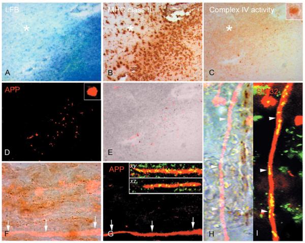
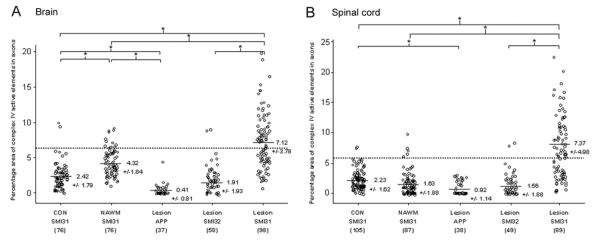
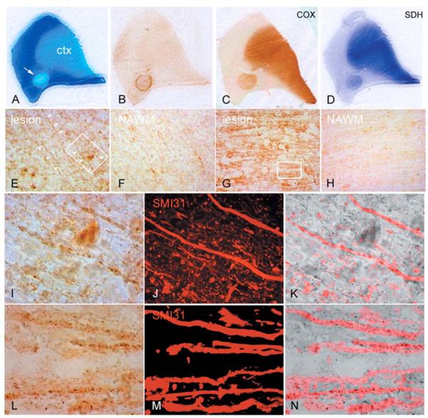
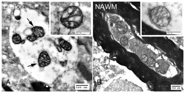
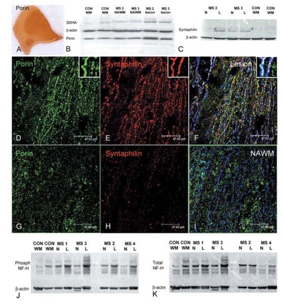
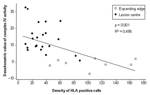
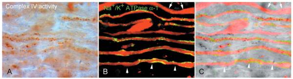
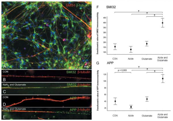
Multiple sclerosis is the most common cause of non-traumatic neurological impairment in young adults. An energy deficient state has been implicated in the degeneration of axons, the pathological correlate of disease progression, in multiple sclerosis. Mitochondria are the most efficient producers of energy and play an important role in calcium homeostasis. We analysed the density and function of mitochondria using immunohistochemistry and histochemistry, respectively, in chronic active and inactive lesions in progressive multiple sclerosis. As shown before in acute pattern III and Balo's lesions, the mitochondrial respiratory chain complex IV activity is reduced despite the presence of mitochondria in demyelinated axons with amyloid precursor protein accumulation, which are predominantly located at the active edge of chronic active lesions. Furthermore, the strong non-phosphorylated neurofilament (SMI32) reactivity was associated with a significant reduction in complex IV activity and mitochondria within demyelinated axons. The complex IV defect associated with axonal injury may be mediated by soluble products of innate immunity, as suggested by an inverse correlation between complex IV activity and macrophage/microglial density in chronic lesions. However, in inactive areas of chronic multiple sclerosis lesions the mitochondrial respiratory chain complex IV activity and mitochondrial mass, judged by porin immunoreactivity, are increased within approximately half of large (>2.5 microm diameter) chronically demyelinated axons compared with large myelinated axons in the brain and spinal cord. The axon-specific mitochondrial docking protein (syntaphilin) and phosphorylated neurofilament-H were increased in chronic lesions. The lack of complex IV activity in a proportion of Na(+)/K(+) ATPase alpha-1 positive demyelinated axons supports axonal dysfunction as a contributor to neurological impairment and disease progression. Furthermore, in vitro studies show that inhibition of complex IV augments glutamate-mediated axonal injury (amyloid precursor protein and SMI32 reactivity). Our findings have important implications for both axonal degeneration and dysfunction during the progressive stage of multiple sclerosis.


Figures

Figure 1
Figure 1
Complex IV activity within injured demyelinated axons. (AE) In the active rim of chronic active multiple sclerosis lesions, identified by the loss of Luxol Fast Blue (A) and presence of MHC class II positive cells (B) in serial sections, the complex IV activity is at an intermediate level compared with normal appearing white matter and relatively inactive lesion centre (C). Surprisingly complex IV activity is increased in the inactive area of chronic lesions. There are a number of ovoid shaped structures containing intense complex IV activity in the active rim (C and insert). When the same serial section used for COX histochemistry (C) is immunofluorescently labelled for amyloid precursor protein (APP) (D), the complex IV active ovoid structures are APP-positive (D, E). However, not all APP reactive ovoids show complex IV activity (red, E). (FI) The APP-positive linear segments of demyelinated axons without terminal ovoids (red in F and G) in chronic lesions are devoid of complex IV activity, brown punctate elements apparent at 100× magnification (F, arrows). The lack of complex IV activity in APP-positive demyelinated axons is not due to the lack of mitochondria as shown by the presence of porin reactive elements (green) in xy and x–z confocal images (G arrows and inserts). In a separate chronic lesion, the immunofluorescent labelling of non-phosphorylated neurofilaments (SMI32) identifies an injured demyelinated axon (red in H and I) which also lacks complex IV active elements (H, arrowheads). The SMI32 (red) reactive axon lacking complex IV active elements contain numerous porin (green) reactive elements (I, arrowheads). x– y and x–z; confocal images of APP and porin immunoreactivity in xy and xz planes. Asterisk indicates inactive area of chronic multiple sclerosis lesion.
Figure 2
Figure 2
Quantitation of complex IV activity within axons in brain and spinal cord. (A, B) The percentage area of complex IV active elements within large (>2.5 μm) axons indicates a significant decrease in complex IV activity within acutely injured APP positive axons in brain (A) and spinal cord (B) multiple sclerosis lesions compared with SMI31 positive myelinated axons. Furthermore, SMI32 positive injured demyelinated axons from inactive areas of chronic lesions contain significantly less complex IV activity compared with SMI31 positive demyelinated axons from the same area. The complex IV activity within SMI31 positive chronically demyelinated axons are significantly greater compared with myelinated axons in normal appearing white matter and control white matter of brain and spinal cord. The SMI31 positive myelinated axons in normal appearing white matter contain significantly greater complex IV activity compared with SMI31 positive myelinated axons in control white matter in brain (A) but not spinal cord (B). The dotted line indicates the upper range of 95% confidence interval based on complex IV activity in myelinated axons from control white matter. The number of large axons analysed is indicated within parentheses. The values are means ± SD of percentage area of complex IV active elements within large axons. *P < 0.001.
Figure 3
Figure 3
Mitochondrial respiratory chain complex IV and complex II activity in chronic multiple sclerosis lesions. (AD) Serial sections of a posterior frontal tissue block containing a chronic active multiple sclerosis lesion (A, arrow), identified by the loss of Luxol Fast Blue (LFB, A) and a rim of inflammatory activity (HLA reactivity, B), shows increased activity of mitochondrial respiratory chain complex IV (C) or cytochrome c oxidase (COX) and complex II (D) or SDH throughout the lesion compared with normal appearing white matter. The complexes IV and II active mitochondria are enriched in the cortex (ctx). (EH) The complex IV active mitochondrial elements are prominent within axons as well as glia in relatively inactive areas of chronic lesions in the brain (E) as well as spinal cord (G) lesions compared with adjacent normal appearing white matter (F and H). (IN) The chronically demyelinated large diameter (>2.5 μm) axons in brain (outlined in E) and spinal cord (outlined in G), identified by SMI31 reactivity (J and M) within chronic lesions, contain intense complex IV active elements (I and L) as evident in the superimposed images (K and N). Most complex IV active elements are punctate (I and L), typical of mitochondria, but others are elongated aligning the longitudinal axis of axons (I).
Figure 4
Figure 4
COX electronmicroscopy. (A,B) Electronmicroscopy of chronic multiple sclerosis lesions following COX histochemistry shows mitochondria with dark cristae and membranes abundant in complex IV activity (A, arrows) within demyelinated axons compared with mitochondria (B, arrowheads) in myelinated axons in normal appearing white matter.
Figure 5
Figure 5
Mass and docking of axonal mitochondria and phosphorylation status of neurofilaments in chronic multiple sclerosis lesions. (AC) Mitochondrial mass, judged by porin immunoreactivity, is increased throughout chronic multiple sclerosis lesions, as apparent in a serial section of the lesion in Fig. 1. Western blots of porin, a voltage gated anion channel expressed on all mitochondria, and a subunit of complex II (SDH70 kDa or SDHA), which is entirely encoded by nuclear DNA, shows a 1.6-fold increase in mitochondrial mass in chronic lesions compared with control white matter (n = 5, P = 0.006). (B). Western blots of syntaphilin, an axon specific mitochondrial docking protein, shows a 3.2- and 1.8-fold increase (n = 4 P = 0.002) in chronic lesions compared with normal appearing white matter and controls, respectively (C). (DI) Porin (green) and syntaphilin (red) reactive elements in triple immunofluorescently labelled confocal images are abundant in chronic lesions (D and E) compared with normal appearing white matter (G and H). The majority of syntaphilin reactive mitochondrial elements are located within SMI31 positive (blue) demyelinated axons (F), whereas myelinated SMI31 positive axons in the normal appearing white matter contain strikingly less porin and syntaphilin reactive elements (I). Interestingly syntaphilin immunoreactivity within demyelinated axons does not always co-localize with porin immunoreactive elements (F). (J, K) Western blots of phosphorylated and total neurofilaments (NF) in chronic lesions show an increase in phosphorylated neurofilament-H in lesions compared with normal appearing white matter and control white matter (J), which is partly due to an increase in total neurofilaments (K). When corrected for β-actin and total neurofilament-H there is a 1.7-fold increase (P = 0.025, n = 6) in phosphorylated neurofilament-H in chronic lesions compared with control white matter. CON WM; control white matter. L = lesion. MS = multiple sclerosis; NAWM or N = normal appearing white matter; NF = neurofilaments.
Figure 6
Figure 6
Inverse correlation between global complex IV activity within multiple sclerosis lesions and the density of microglia/macrophages. When the global complex IV activity was densitometrically determined in 20× images, there is a significant inverse correlation (P = 0.001) between complex IV activity and the density of HLA reactive microglia and phagocytic macrophages. Furthermore, complex IV activity in the active rims or expanding edge of chronic active lesions (unfilled circles) is significantly lower (P < 0.001) compared with the inactive areas of chronic lesions (filled circles). The cell density is per 20× field. The densitometric values are corrected for background variation using normal appearing white matter.
Figure 7
Figure 7
The complex IV dysfunction involves chronically demyelinated axons with Na+/K+ ATPase α-1 reactivity. (AC) As a proportion of chronically demyelinated axons lacks Na+/K+ ATPase (Young et al., 2008), necessary for sodium extrusion from the axon, complex IV activity was determined within Na+/K+ ATPase α-1 positive chronically demyelinated axons. There are Na+/K+ ATPase α-1 positive demyelinated axons, identified with total neurofilaments, in inactive areas of chronic lesions showing a lack of complex IV active elements (arrowheads). Furthermore, there are axons with bulbous expansions lacking complex IV activity as well as axons lacking both Na+/K+ ATPase α-1 and complex IV activity (arrows).
Figure 8
Figure 8
Complex IV inhibition augments glutamate-mediated axonal injury, in vitro. (AE) The axons in control neuron cultures, differentiated from mouse embryonic stem cells, in vitro, express both β-tubulin (A, green) as well as phosphorylated neurofilaments (SMI31 in a, red). The neuronal cultures were exposed to sublethal concentrations and durations of sodium azide (10 μM sodium azide for 120 min), a specific complex IV inhibitor, or glutamate (100 μM glutamate for 60 min) alone as well as sodium azide and glutamate sequentially (addition of 100 μM glutamate to the second 1 h of the 2 h 10 μM sodium azide exposure). Within β-tubulin positive unmyelinated axons not exposed to sodium azide or glutamate (B and D, red) there is minimal SMI32 (non-phosphorylated neurofilaments) and APP (amyloid precursor protein) reactivity (B and D, green). In contrast, neuron cultures exposed to sublethal concentrations of sodium azide and glutamate show abundant SMI32 (C, green) and APP (E, green) reactivity within β-tubulin positive (C and E, red) unmyelinated axons. (F, G) When acute injury of unmyelinated axons, in vitro, was densitometrically analysed the 10 μM sodium azide, which reduced complex IV activity by 36%, led to a significant decrease in APP immunoreactivity in β-tubulin positive axons (F). When the neuronal cultures were sequentially exposed to sodium azide there is a striking synergistic increase in axonal injury, judged by both SMI32 (F) and APP (G) immunoreactivity. *P < 0.001. Two hundred axons were analysed in four separate experiments for each group. CON = control; NaN3 = sodium azide.

References

    1. Ames A., 3rd CNS energy metabolism as related to function. Brain Res Brain Res Rev. 2000;34:42–68. - PubMed
    1. Amiri M, Hollenbeck PJ. Mitochondrial biogenesis in the axons of vertebrate peripheral neurons. Dev Neurobiol. 2008;68:1348–61. - PMC - PubMed
    1. Andrews H, White K, Thomson C, Edgar J, Bates D, Griffiths I, et al. Increased axonal mitochondrial activity as an adaptation to myelin deficiency in the Shiverer mouse. J Neurosci Res. 2006;83:1533–9. - PubMed
    1. Andreyev AY, Kushnareva YE, Starkov AA. Mitochondrial metabolism of reactive oxygen species. Biochemistry. 2005;70:200–14. - PubMed
    1. Barron MJ, Griffiths P, Turnbull DM, Bates D, Nichols P. The distributions of mitochondria and sodium channels reflect the specific energy requirements and conduction properties of the human optic nerve head. Br J Ophthalmol. 2004;88:286–90. - PMC - PubMed

Publication types

MeSH terms