. 2009 Apr;132(Pt 4):889-902.
doi: 10.1093/brain/awp029. Epub 2009 Mar 17.

Obesity induces functional astrocytic leptin receptors in hypothalamus

Affiliations

Obesity induces functional astrocytic leptin receptors in hypothalamus

Hung Hsuchou et al. Brain. 2009 Apr.

Abstract

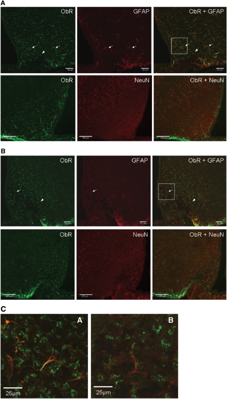
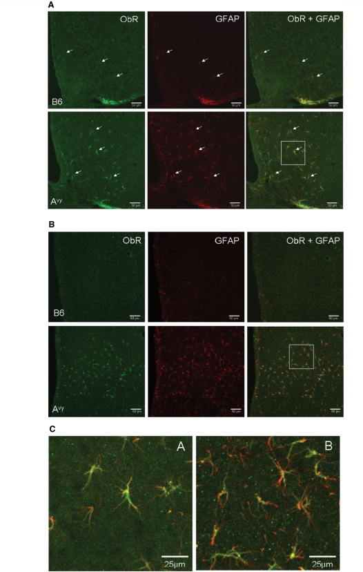
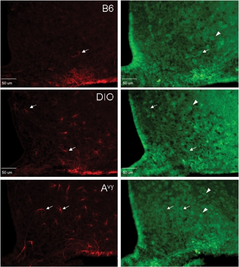
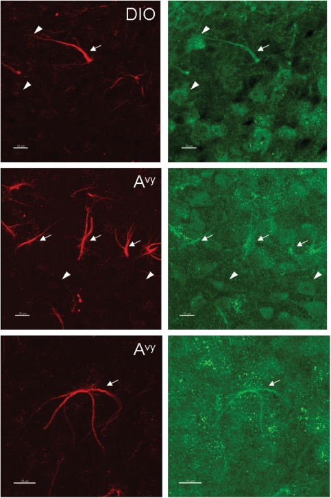
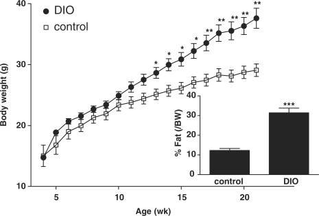
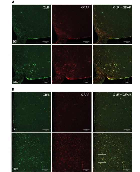
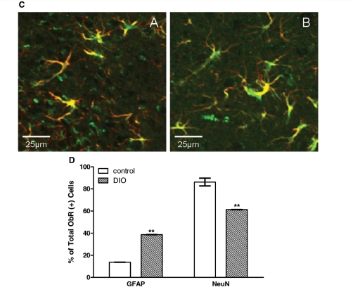
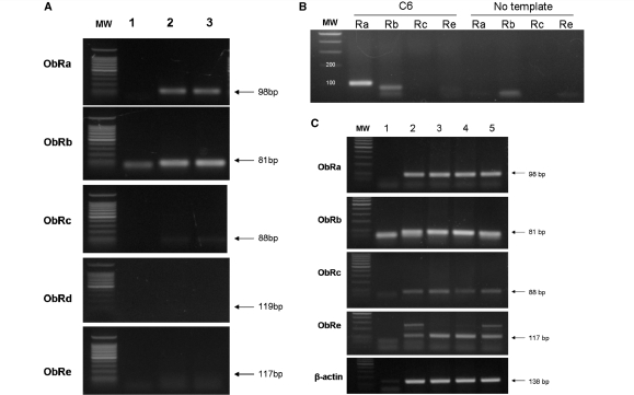
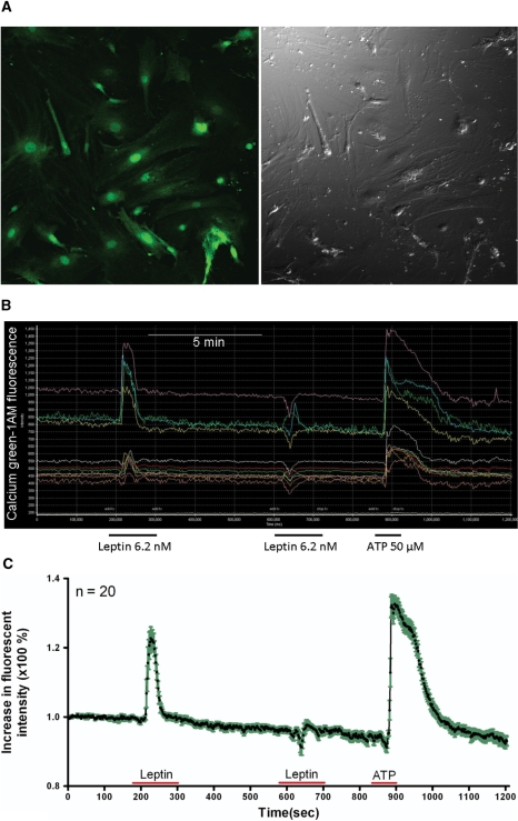
The possible role of astrocytes in the regulation of feeding has been overlooked. It is well-established that the endothelial cells constituting the blood-brain barrier transport leptin from blood to brain and that hypothalamic neurons respond to leptin to induce anorexic signaling. However, few studies have addressed the role of astrocytes in either leptin transport or cellular activation. We recently showed that the obese agouti viable yellow mouse has prominent astrocytic expression of the leptin receptor. In this study, we test the hypothesis that diet-induced obesity increases astrocytic leptin receptor expression and function in the hypothalamus. Double-labelling immunohistochemistry and confocal microscopic analysis showed that all astrocytes in the hypothalamus express leptin receptors. In adult obese mice, 2 months after being placed on a high-fat diet, there was a striking increase of leptin receptor (+) astrocytes, most prominent in the dorsomedial hypothalamus and arcuate nucleus. Agouti viable yellow mice with their adult-onset obesity showed similar changes, but the increase of leptin receptor (+) astrocytes was barely seen in ob/ob or db/db mice with their early-onset obesity and defective leptin systems. The marked leptin receptor protein expression in the astrocytes, shown with several antibodies against different receptor epitopes, was supported by RT-PCR detection of leptin receptor-a and -b mRNAs in primary hypothalamic astrocytes. Unexpectedly, the protein expression of GFAP, a marker of astrocytes, was also increased in adult-onset obesity. Real-time confocal imaging showed that leptin caused a robust increase of calcium signalling in primary astrocytes from the hypothalamus, confirming their functionality. The results indicate that metabolic changes in obese mice can rapidly alter leptin receptor expression and astrocytic activity, and that leptin receptor is responsible for leptin-induced calcium signalling in astrocytes. This novel and clinically relevant finding opens new avenues in astrocyte biology.


Figures

Figure 1
Figure 1
Expression of ObR in arcuate nucleus of different mouse models shown by immunostaining with M18 antibody. Astrocytes were immunostained with anti-GFAP (red, arrows), whereas neurons were immunostained with anti-NeuN (red, arrow heads). Yellow colouration indicates co-localization. (A) ob/ob mouse; (B) db/db mouse (confocal image, scale bar = 50 µm for GFAP; scale bar = 100 µm for NeuN). (C) Higher magnification of the co-localization of ObR and GFAP from the demarcated areas in A and B. Scale bar: 25 μm.
Figure 2
Figure 2
Expression of ObR in the arcuate nucleus (A) and dorsomedial hypothalamus (B) shown by immunostaining with the K20 antibody. In both regions, there were more ObR (+) astrocytes (arrows) in Avy than in age-matched B6 mice (confocal image, scale bar = 50 µm). (C) Higher magnification of the co-localization of ObR and GFAP from the demarcated areas in (A and B).
Figure 3
Figure 3
Expression of ObR in the arcuate nucleus of B6 (top), diet-induced obesity (DIO) (middle) and Avy (bottom) mice shown by double immunostaining with M-18 (red) and CH14104 (green) antibodies. (A) M-18 immunoreactivity was mainly seen in astrocytes (arrows). CH14104 antibody stained mostly neurons (arrow heads). In comparison with the lean B6 control, the diet-induced obesityB6 mice and the Avy mice on a B6 background showed a greater number of ObR(+) astrocytes seen with the M18 antibody (epifluorescence image, scale bar = 50 µm). (B) Higher magnification images showing that astrocytes can be stained with both M-18 and CH14104 antibodies, the former providing a stronger signal with a crisper background (confocal image, scale bar = 10 µm).
Figure 3
Figure 3
Expression of ObR in the arcuate nucleus of B6 (top), diet-induced obesity (DIO) (middle) and Avy (bottom) mice shown by double immunostaining with M-18 (red) and CH14104 (green) antibodies. (A) M-18 immunoreactivity was mainly seen in astrocytes (arrows). CH14104 antibody stained mostly neurons (arrow heads). In comparison with the lean B6 control, the diet-induced obesityB6 mice and the Avy mice on a B6 background showed a greater number of ObR(+) astrocytes seen with the M18 antibody (epifluorescence image, scale bar = 50 µm). (B) Higher magnification images showing that astrocytes can be stained with both M-18 and CH14104 antibodies, the former providing a stronger signal with a crisper background (confocal image, scale bar = 10 µm).
Figure 4
Figure 4
Obesity phenotype of the diet-induced obesity mice (n = 9), shown by more rapid increase of body weight over time and higher percentage of body fat at 8.5 weeks in comparison with their former littermates fed with regular rodent chow (n = 7). *P < 0.05; **P < 0.01; ***P < 0.005.
Figure 5
Figure 5
Diet-induced obesity increased ObR (+) astrocytes in the arcuate nucleus (A) and to an even greater extent in the dorsomedial hypothalamic nucleus (B) (confocal image, scale bar = 100 µm). (C) Higher magnification of the co-localization of ObR and GFAP from the demarcated areas above in diet-induced obesity. Scale bar: 25 μm. (D) Percent of ObR (+) cells that were also GFAP or NeuN (+); the diet-induced obesity group had significantly more ObR (+) astrocytes in the arcuate nucleus than the controls, with corresponding reduction of ObR (+) neurons. **P < 0.01.
Figure 5
Figure 5
Diet-induced obesity increased ObR (+) astrocytes in the arcuate nucleus (A) and to an even greater extent in the dorsomedial hypothalamic nucleus (B) (confocal image, scale bar = 100 µm). (C) Higher magnification of the co-localization of ObR and GFAP from the demarcated areas above in diet-induced obesity. Scale bar: 25 μm. (D) Percent of ObR (+) cells that were also GFAP or NeuN (+); the diet-induced obesity group had significantly more ObR (+) astrocytes in the arcuate nucleus than the controls, with corresponding reduction of ObR (+) neurons. **P < 0.01.
Figure 6
Figure 6
RT–PCR analysis shows that astrocytes expressed mainly ObRa (98 bp) and ObRb (81 bp) mRNA. (A) Primary astrocytes from 1-week-old mouse pups. Lane 1: no template control; lane 2: primary astrocytes from mouse hypothalamus; lane 3: primary astrocytes from mouse striatum. (B) C6 astrocytoma cells with rat-specific primers. (C) In contrast, mouse hippocampus had not only ObRa and ObRb, but also ObRc and ObRe. Lane 1: negative control; lanes 2–5: hippocampal tissue from different mice.
Figure 7
Figure 7
Leptin-induced calcium signalling in primary astrocytes from mouse hypothalamus. (A) A field of astrocytes on glass coverslips after calcium green-1AM loading. (B) Time-lapse image showing that all cells responded to leptin and ATP. (C) Time-lapse image showing average amplitude of the calcium response (n = 20). Green area surrounding the line shows SD of the response from individual cells.

Similar articles

Cited by

References

    1. Abbott NJ. Dynamics of CNS barriers: evolution, differentiation, and modulation. Cell Mol Neurobiol. 2005;25:5–23. - PMC - PubMed
    1. Abbott NJ. Cellular compositions of the blood-brain barrier (BBB) In: Kastin AJ, Pan W, editors. The Henry Stewart Talk series: the blood-brain barrier. London: Henry Stewart Talks; 2008.
    1. Abbott NJ, Ronnback L, Hansson E. Astrocyte-endothelial interactions at the blood-brain barrier. Nat Rev Neurosci. 2006;7:41–53. - PubMed
    1. Agulhon C, Petravicz J, McMullen AB, Sweger EJ, Minton SK, Taves SR, et al. What is the role of astrocyte calcium in neurophysiology? Neuron. 2008;59:932–46. - PMC - PubMed
    1. Ahmed M, Shaban Z, Yamaji D, Okamatsu-Ogura Y, Soliman M, Abd EM, et al. Induction of proinflammatory cytokines and caspase-1 by leptin in monocyte/macrophages from holstein cows. J Vet Med Sci. 2007;69:509–14. - PubMed

Publication types