. 2009 Jun;296(6):E1354-62.
doi: 10.1152/ajpendo.90836.2008. Epub 2009 Mar 17.

Role for malic enzyme, pyruvate carboxylation, and mitochondrial malate import in glucose-stimulated insulin secretion

Affiliations

Role for malic enzyme, pyruvate carboxylation, and mitochondrial malate import in glucose-stimulated insulin secretion

Emma Heart et al. Am J Physiol Endocrinol Metab. 2009 Jun.

Abstract

Pyruvate cycling has been implicated in glucose-stimulated insulin secretion (GSIS) from pancreatic beta-cells. The operation of some pyruvate cycling pathways is proposed to necessitate malate export from the mitochondria and NADP(+)-dependent decarboxylation of malate to pyruvate by cytosolic malic enzyme (ME1). Evidence in favor of and against a role of ME1 in GSIS has been presented by others using small interfering RNA-mediated suppression of ME1. ME1 was also proposed to account for methyl succinate-stimulated insulin secretion (MSSIS), which has been hypothesized to occur via succinate entry into the mitochondria in exchange for malate and subsequent malate conversion to pyruvate. In contrast to rat, mouse beta-cells lack ME1 activity, which was suggested to explain their lack of MSSIS. However, this hypothesis was not tested. In this report, we demonstrate that although adenoviral-mediated overexpression of ME1 greatly augments GSIS in rat insulinoma INS-1 832/13 cells, it does not restore MSSIS, nor does it significantly affect GSIS in mouse islets. The increase in GSIS following ME1 overexpression in INS-1 832/13 cells did not alter the ATP-to-ADP ratio but was accompanied by increases in malate and citrate levels. Increased malate and citrate levels were also observed after INS-1 832/13 cells were treated with the malate-permeable analog dimethyl malate. These data suggest that although ME1 overexpression augments anaplerosis and GSIS in INS-1 832/13 cells, it is not likely involved in MSSIS and GSIS in pancreatic islets.


Figures
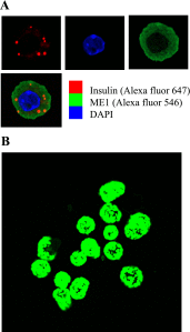
Fig. 1.
Fig. 1.
Cytosolic malic enzyme (ME1) expression in a single mouse β-cell and an intact islet. A: intracellular localization of human ME1 (hME1) protein was determined using an anti-hME1 antibody and Alexa fluor 546. Insulin was detected using anti-insulin antibody and Alexa fluor 647. Nuclei were detected using 4′,6-diamidino-2-phenylindole (DAPI). B: a group of mouse islets infected with Ad-ME1-GFP. Strength of the green fluorescent protein (GFP) signal (excitation at 488/550 nm) was used to determine infection efficiency.
Fig. 2.
Fig. 2.
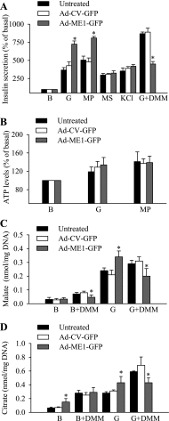
Effect of ME1 overexpression on insulin secretion, ATP, and metabolite levels in INS-1 832/13 cells. INS-1 832/13 cells were left untreated or transduced with Ad-CV-GFP or Ad-ME1-GFP in the absence or presence of Ad-CytoLuc at 50 multiplicity of infection. A: after 2 h of preincubation in the presence of 2 mM glucose (G), insulin secretion in response to secretory fuels at 10 mM or KCl was measured in static incubation over a 30-min period. Basal secretion (B) at 2 mM glucose was 20.5 ± 2.2, 23.2 ± 3.1, and 22 ± 2.7 ng insulin·mg protein−1·h−1 in untreated, Ad-CV-GFP-treated, and Ad-ME1-GFP-treated cells, respectively. Nonstimulatory 2 mM glucose was present during incubation with methyl pyruvate (MP), methyl succinate (MS), and KCl; stimulatory 10 mM glucose was present during incubation with dimethyl malate (DMM). Values are means ± SE from 5 independent experiments. B: ATP levels, measured in real time as relative light output, were determined in a population of ∼0.5 × 10⁶ live cells using the luciferin-luciferase reaction in response to 10 mM glucose and MP. Values are means ± SE from 4 independent measurements. C and D: malate and citrate levels were determined in mitochondrial fractions using tandem mass spectrometry (LC-MS/MS). *P < 0.05 vs. Ad-CV-GFP.
Fig. 3.
Fig. 3.
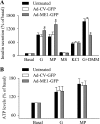
Effect of ME1 overexpression on insulin secretion and ATP levels in mouse islets. Isolated mouse islets were left untreated or transduced with Ad-CV-GFP or Ad-ME1-GFP in the absence (secretion, A) or presence (ATP levels, B) of Ad-CytoLuc at 50 multiplicity of infection. A: after 2 h of preincubation in the presence of 4 mM glucose, insulin secretion was measured in response to secretory fuels at 10 mM or KCl in static incubation over a 30-min period. Basal secretion at 4 mM glucose was 3.2 ± 0.4, 3.7 ± 0.45, and 3.6 ± 0.5 ng insulin·10 islets−1·h−1 in untreated, Ad-CV-GFP-treated, and Ad-ME1-GFP-treated islets, respectively. Nonstimulatory 4 mM glucose was present during incubation with MP, MS, and KCl. Stimulatory glucose (10 mM) was present during incubation with DMM. Values are means ± SE from 3–4 independent experiments. B: ATP production, measured as relative light output, was determined in a population of ∼3.5 × 10⁶ live islets cells (obtained by dispersion of whole islets) using the luciferin-luciferase reaction in response to stimulatory (10 mM) concentration of fuels. Values are means ± SE from 3 independent experiments. *P < 0.05 vs. Ad-CV-GFP.
Fig. 4.
Fig. 4.
MS-mediated metabolic response is not contingent on ME1 activity. A and B: a single mouse β-cell Ca2+ response in cells treated with Ad-ME1-GFP and in control cells. Addition of secretory fuels at 10 mM is indicated by arrows. Fura Red fluorescence is expressed as relative fluorescence units (F1/F0, where F0 is mean baseline fluorescence). Note that Fura Red fluorescence intensity decreases upon Ca2+ binding (15). C: MIN-6 cells do not respond to MS, despite ME1 activity. After a 2-h preincubation in the presence of 2 mM glucose, MIN-6 cells were exposed to secretory fuels at 10 mM. Control cells were treated with 2 mM glucose (basal secretion). Insulin secretion was measured in static incubation over a 30-min period. Basal secretion was 56 ± 5.5 insulin·mg protein−1·h−1. NADP+-dependent malate dehydrogenase activities were measured in the soluble protein fraction. Values are means ± SE from 4–5 independent experiments.
Fig. 5.
Fig. 5.
Differential effect of MS on O2 consumption in mouse and rat islets. A: MS does not stimulate a sustained increase in O2 consumption in mouse islets. B: MS elicits a sustained increase in O2 consumption in rat islets. Addition of fuels (10 mM) is indicated by arrows. Each trace is representative of 4–6 independent measurements. C: ME1 overexpression does not restore MS-mediated O2 consumption in mouse islets. Values are means ± SE from 3 independent experiments.
Fig. 6.
Fig. 6.
DMM enhances insulin secretion: role of malate import into mitochondria. At initially high levels of cytosolic pyruvate (glycolysis) and low levels of cytosolic malate, the ME1-catalyzed reaction proceeds in the direction of malate formation and cytosolic malate enters mitochondria, resulting in increased output of citrate or isocitrate and enhanced insulin secretion. However, when the level of cytosolic malate becomes high (e.g., after DMM application), the ME1-dependent reaction functions in reverse, favoring pyruvate formation (dotted line), resulting in decreased citrate/isocitrate output and insulin secretion. Under the second condition, increased ME1 activity (ME1 overexpression) compared with cells with lower endogeneous (INS-1 832/13) or low or absent (mouse islets) ME-1 activity facilitates removal of cytosolic malate and removes DMM potentiation of glucose-stimulated insulin secretion (GSIS) via decreased citrate/isocitrate output. OAA, oxaloacetate; TCC, tricarboxylate carrier; ICDc, cytosolic isocitrate dehydrogenase; α-KG, α-ketoglutarate; PC, pyruvate carboxylase; PDH, pyruvate dehydrogenase; SCI, secretory coupling intermediates.

Similar articles

Cited by

References

    1. Antinozzi PA, Segall L, Prentki M, McGarry JD, Newgard CB. Molecular or pharmacologic perturbation of the link between glucose and lipid metabolism is without effect on glucose-stimulated insulin secretion. A re-evaluation of the long-chain acyl-CoA hypothesis. J Biol Chem 273: 16146–16154, 1998. - PubMed
    1. Ashcroft SJ, Randle PJ. Enzymes of glucose metabolism in normal mouse pancreatic islets. Biochem J 119: 5–15, 1970. - PMC - PubMed
    1. Berry MN The function of energy-dependent redox reactions in cell metabolism. FEBS Lett 117 Suppl: K106–K120, 1980. - PubMed
    1. Berry MN, Fanning DC, Grivell AR, Lewis SJ, Farrington CJ, Wallace PG. Evidence for several separate functional pools of NAD(H) within the cytoplasmic compartment of the hepatocyte. Biochem Soc Trans 8: 570, 1980. - PubMed
    1. Boucher A, Lu D, Burgess SC, Telemaque-Potts S, Jensen MV, Mulder H, Wang MY, Unger RH, Sherry AD, Newgard CB. Biochemical mechanism of lipid-induced impairment of glucose-stimulated insulin secretion and reversal with a malate analogue. J Biol Chem 279: 27263–27271, 2004. - PubMed

Publication types

MeSH terms