. 2009 Jun;85(6):957-65.
doi: 10.1189/jlb.0908575. Epub 2009 Mar 17.

Lnk inhibits myeloproliferative disorder-associated JAK2 mutant, JAK2V617F

Affiliations

Lnk inhibits myeloproliferative disorder-associated JAK2 mutant, JAK2V617F

Sigal Gery et al. J Leukoc Biol. 2009 Jun.

Abstract

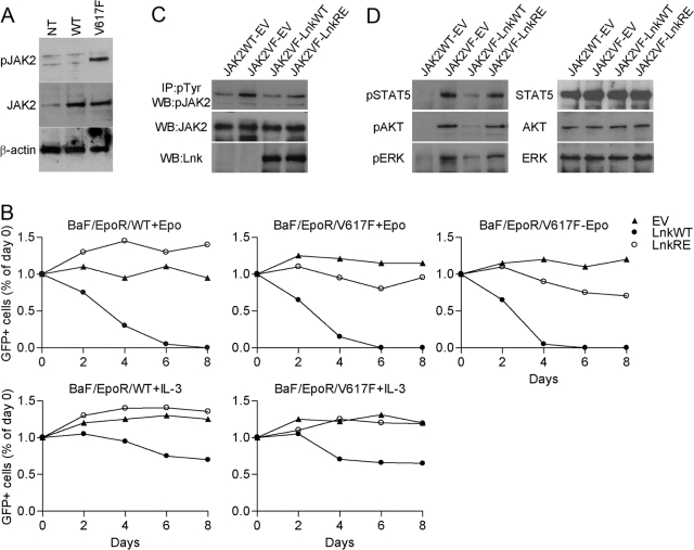
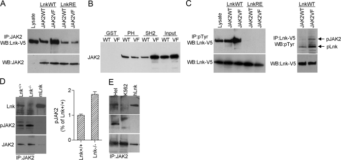
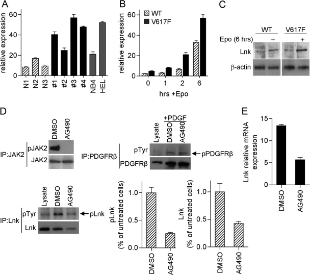
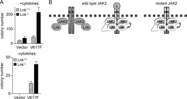
The JAK2 mutation JAK2V617F is found frequently in patients with myeloproliferative disorders (MPD) and transforms hematopoietic cells to cytokine-independent proliferation when expressed with specific cytokine receptors. The Src homology 2 (SH2) and pleckstrin homology (PH) domain-containing adaptor protein Lnk (SH2B3) is a negative regulator of hematopoietic cytokine signaling. Here, we show that Lnk is a potent inhibitor of JAK2V617F constitutive activity. Lnk down-regulates JAK2V617F-mediated signaling and transformation in hematopoietic Ba/F3-erythropoietin receptor cells. Furthermore, in CFU assays, Lnk-deficient murine bone marrow cells are significantly more sensitive to transformation by JAK2V617F than wild-type (WT) cells. Lnk, through its SH2 and PH domains, interacts with WT and mutant JAK2 and is phosphorylated by constitutively activated JAK2V617F. Finally, we found that Lnk levels are high in CD34(+) hematopoietic progenitors from MPD patients and that Lnk expression is induced following JAK2 activation. Our data suggest that JAK2V617F is susceptible to endogenous negative-feedback regulation, providing new insights into the molecular pathogenesis of MPD.


Figures

Fig. 1.
Fig. 1.
Lnk inhibits proliferation of hematopoietic cells expressing JAK2V617F. (A) Western blot analysis of JAK2 total protein and phosphorylation levels in the Ba/F3-EpoR cells stably expressing JAK2WT (BaF/EpoR/WT) or JAK2V617F (BaF/EpoR/V617F). Cells were depleted of cytokines for 16 h. NT, Nontransfected Ba/F3-EpoR cells. (B) BaF/EpoR/WT and BaF/EpoR/V617F cells were transduced with MSCV-IRES-GFP (MIG) empty vector (EV), MIG WT Lnk (LnkWT), or MIG SH2 mutant Lnk (LnkRE). Two days later (Day 0), the percent of transfected cells was analyzed by measuring GFP (considered as 100%). Subsequently, GFP expression was measured and calculated relative to that of Day 0. Epo (10 U/ml) and IL-3 (10 ng/ml) were added as indicted. Results are representative of three independent experiments. (C and D) BaF/EpoR/WT and BaF/EpoR/V617F cells were transduced with the MIG vectors and cultured for 2 days with (BaF/EpoR/WT) or without Epo (BaF/EpoR/V617F), after which, both cell lines were incubated without Epo for 16 h. FACS-sorted GFP-positive cells were lysed. (C) Levels of pJAK2 were analyzed by immunoprecipitation (IP) with pTyr antibody, followed by Western blot (WB) with pJAK2 antibody. Levels of total JAK2 and Lnk were analyzed by Western blot. (D) Western blot analysis of phosphorylation and total levels of the indicated proteins. Experiments were repeated three times with similar results.
Fig. 2.
Fig. 2.
Lnk interacts with JAK2WT and JAK2V617F. (A–C) 293T cells were cotransfected with combinations of JAKWT (JAK2WT), JAK2V617F (JAK2VF), WT Lnk-V5 (LnkWT), or SH2 mutant Lnk-V5 (LnkRE) as indicated. Lysates were immunoprecipitated with JAK2, pTyr, or V5 antibodies and analyzed by Western blot as indicated (A and C, upper panels). JAK2 and Lnk levels in the lysates were analyzed by Western blot with JAK2 (A) and V5 (C) antibodies (lower panels). Lysate from 293T-Lnk-V5-transfected cells was used as control for molecular size (A and C, lysate, first lane). (B) Lysates from 293T cells transfected with JAK2WT or JAK2V617F were incubated with GST, GST-Lnk PH (PH), or GST-Lnk-SH2 (SH2) fusion proteins. GST-protein complexes were analyzed by Western blot with JAK2 antibody. Input represents one-tenth of the lysate used for the pull-downs. (D) BM cells from Lnk+/+ and Lnk−/− mice were starved in RPMI 1640 for 4 h and stimulated with 20 ng/ml SCF, 10 U/ml Epo, and 20 ng/ml Tpo for 30 min. Lysates were immunoprecipitated with JAK2 antibody. Bar graph shows the mean ± sd of pJAK2 from two experiments. Results are expressed as a relative percentage compared with pJAK2 in Lnk+/+ cells. (E) Lysates from Hel and K562 cells were immunoprecipitated with JAK2 antibody. (E and D) Blots were probed with the indicated antibodies. Lysates from 293T Lnk-transfected cells were used to control for molecular size. mLnk, Murine Lnk; hLnk, human Lnk. Shown are representative results from two independent experiments.
Fig. 3.
Fig. 3.
High levels of Lnk are associated with MPD. (A) Lnk expression was measured by real-time PCR in CD34+ hematopoietic progenitor cells from MPD patients (JAK2V617F-negative #1 and #2; JAK2V617F-positive #3 and #4), as well as normal donors (N1–N3) and cell lines NB4 (JAK2WT) and Hel (JAK2V617F). Relative Lnk mRNA levels are expressed in arbitrary units as a ratio of Lnk transcripts:18S transcripts. Data represent the mean ± sd of triplicate samples. (B and C) Real-time PCR (B) and Western blot (C) analysis of Lnk expression in Ba/F3 cells stably expressing EpoR and JAK2WT (WT) or JAK2V617F (V617F). Cells were depleted of cytokines for 16 h and then stimulated with Epo (10 U/ml) for the indicated times. Experiments were repeated three times. (D and E) Hel cells were treated for 4 h with JAK2 inhibitor (AG490, 50 μM) or vehicle alone (DMSO). (D) pJAK2 was analyzed by immunoprecipitation with JAK2 antibody followed by Western blot with pJAK2 antibody; JAK2 protein level was analyzed by Western blot. pLnk was analyzed by immunoprecipitation with pTyr antibody followed by Western blot with Lnk antibody; total Lnk levels were analyzed by Western blot. Bar graphs show the mean ± sd of pLnk and Lnk from three experiments. Results are expressed as a relative percentage compared with untreated cells. As a negative control, pPDGFRβ was analyzed in cells stimulated with PDGF-BB (20 ng/ml) for the last 5 min of AG490/DMSO treatment. Lysates from 293T Lnk- and PDGFRβ-transfected cells were used to control for molecular size. (E) Lnk mRNA levels were measured by real-time PCR. Relative Lnk mRNA levels are expressed in arbitrary units as a ratio of Lnk transcripts:18S transcripts. Data represent the mean ± sd of triplicate samples. Shown are representative results from three experiments.
Fig. 4.
Fig. 4.
Lnk inhibits JAK2V617F-induced transformation in primary murine cells. (A) BM cells from Lnk+/+ and Lnk−/− mice (three of each) were transduced with MIG vector alone or MIG-JAK2V617F and sorted for GFP expression. Methylcellulose CFU assays were performed in the absence or presence of cytokines. Total colony numbers (including CFU-E, BFU-E, CFU-M, CFU-GM, and CFU-G) were counted on Day 6 (with cytokines) and Day 12 (without cytokines). Data represent the mean ± sd of triplicate samples and are representative of two experiments. (B) A model showing Lnk interactions with WT and mutant JAK2. In the absence of ligand (L), Lnk interacts weakly with non-pWT JAK2. Following ligand stimulation, activated JAK2 induces signaling by phosphorylating downstream positive and negative regulators, including Lnk. Consequently, Lnk strongly binds to pTyr in JAK2 and the receptor (R), thereby attenuating the activated receptor/JAK2 complex. In cells expressing mutant JAK2, downstream positive and negative regulators are constitutively activated, leading to an overall enhanced signaling. Dark gray shapes, Nonphosphorylated proteins; bright shapes, phosphorylated proteins.

References

    1. Parganas E, Wang D, Stravopodis D, Topham D J, Marine J C, Teglund S, Vanin E F, Bodner S, Colamonici O R, van Deursen J M, Grosveld G, Ihle J N. Jak2 is essential for signaling through a variety of cytokine receptors. Cell. 1998;93:385–395. - PubMed
    1. Campbell P J, Green A R. The myeloproliferative disorders. N Engl J Med. 2006;355:2452–2466. - PubMed
    1. Levine R L, Pardanani A, Tefferi A, Gilliland D G. Role of JAK2 in the pathogenesis and therapy of myeloproliferative disorders. Nat Rev Cancer. 2007;7:673–683. - PubMed
    1. Baxter E J, Scott L M, Campbell P J, East C, Fourouclas N, Swanton S, Vassiliou G S, Bench A J, Boyd E M, Curtin N, Scott M A, Erber W N, Green A R. Cancer Genome Project Acquired mutation of the tyrosine kinase JAK2 in human myeloproliferative disorders. Lancet. 2005;365:1054–1061. - PubMed
    1. James C, Ugo V, Le Couedic J P, Staerk J, Delhommeau F, Lacout C, Garcon L, Raslova H, Berger R, Bennaceur-Griscelli A, Villeval J L, Constantinescu S N, Casadevall N, Vainchenker W. A unique clonal JAK2 mutation leading to constitutive signaling causes polycythemia vera. Nature. 2005;434:1144–1148. - PubMed

Publication types

MeSH terms