. 2009 Apr;110(4):813-25.
doi: 10.1097/ALN.0b013e31819b602b.

Inhibition of p75 neurotrophin receptor attenuates isoflurane-mediated neuronal apoptosis in the neonatal central nervous system

Affiliations

Inhibition of p75 neurotrophin receptor attenuates isoflurane-mediated neuronal apoptosis in the neonatal central nervous system

Brian P Head et al. Anesthesiology. 2009 Apr.

Abstract

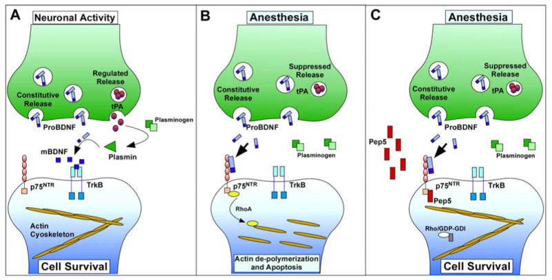
Background: Exposure to anesthetics during synaptogenesis results in apoptosis and subsequent cognitive dysfunction in adulthood. Probrain-derived neurotrophic factor (proBDNF) is involved in synaptogenesis and can induce neuronal apoptosis via p75 neurotrophic receptors (p75). proBDNF is cleaved into mature BDNF (mBDNF) by plasmin, a protease converted from plasminogen by tissue plasminogen activator (tPA) that is released with neuronal activity; mBDNF supports survival and stabilizes synapses through tropomyosin receptor kinase B. The authors hypothesized that anesthetics suppress tPA release from neurons, enhance p75 signaling, and reduce synapses, resulting in apoptosis.

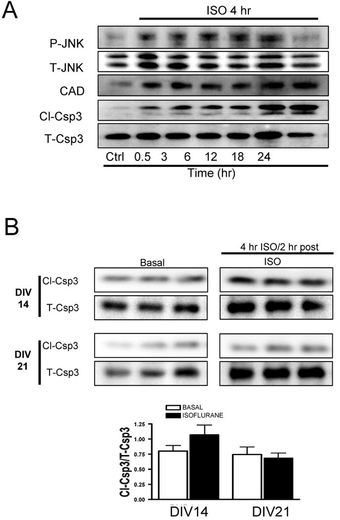
Methods: Primary neurons (DIV5) and postnatal day 5-7 (PND5-7) mice were exposed to isoflurane (1.4%, 4 h) in 5% CO2, 95% air. Apoptosis was assessed by cleaved caspase-3 (Cl-Csp3) immunoblot and immunofluorescence microscopy. Dendritic spine changes were evaluated with the neuronal spine marker, drebrin. Changes in synapses in PND5-7 mouse hippocampi were assessed by electron microscopy. Primary neurons were exposed to tPA, plasmin, or pharmacologic inhibitors of p75 (Fc-p75 or TAT-Pep5) 15 min before isoflurane. TAT-Pep5 was administered by intraperitoneal injection to PND5-7 mice 15 min before isoflurane.

Results: Exposure of neurons in vitro (DIV5) to isoflurane decreased tPA in the culture medium, reduced drebrin expression (marker of dendritic filopodial spines), and enhanced Cl-Csp3. tPA, plasmin, or TAT-Pep5 stabilized dendritic filopodial spines and decreased Cl-Csp3 in neurons. TAT-Pep5 blocked isoflurane-mediated increase in Cl-Csp3 and reduced synapses in PND5-7 mouse hippocampi.

Conclusion: tPA, plasmin, or p75 inhibition blocked isoflurane-mediated reduction in dendritic filopodial spines and neuronal apoptosis in vitro. Isoflurane reduced synapses and enhanced Cl-Csp3 in the hippocampus of PND5-7 mice, the latter effect being mitigated by p75 inhibition in vivo. These data support the hypothesis that isoflurane neurotoxicity in the developing rodent brain is mediated by reduced synaptic tPA release and enhanced proBDNF/p75-mediated apoptosis.


Figures

Fig. 1
Fig. 1
Isoflurane exposure increases cell death in DIV5, but not DIV14 or DIV21 in vitro. Primary neurons (5 days in vitro - DIV) were exposed to 1.4% isoflurane for 4 h and neurons were lysed at various time intervals post exposure. The pro-apoptotic proteins, P-JNK, CAD, and cleaved caspase-3 (Cl-Csp3), were enhanced at 0.5, 3, 6, 12, 18, and 24 h (with the exception of P-JNK which returned to basal levels prior to 24 hr) post isoflurane exposure as shown in 1A. Primary neonatal neurons (DIV14 and DIV21) were exposed to isoflurane and representative immunoblots are shown in Fig 1B. Isoflurane slightly increased (not significant) apoptosis in DIV14 (p=0.2191, n=3) but had no effect on DIV21 (p=0.6821, n=3) neonatal neurons. In contrast to DIV5 neurons, these findings indicate that DIV14 and DIV21 neurons are not vulnerable to isoflurane-induced apoptosis. Error bars, standard error of the mean (s.e.m.).
Fig. 2
Fig. 2
Tissue plasminogen activator (tPA) decreases isoflurane-induced cell death in DIV5 primary neurons. Primary neurons (5 days in vitro - DIV) were exposed to 1.4% isoflurane for 4 h and the media was frozen 2 h post exposure at -80°C prior to tPA ELISA. The media from neurons exposed to isoflurane (4 h - 1.4%; 2 h post) contained significantly (*n=6, p=0.001) less tPA compared to media from non-exposed cells (∼45% decrease; 1.40 ng/ml-BASAL vs 0.77 ng/ml-isoflurane) as shown in 2A. Neurons were pre-treated with increasing doses of tPA (0.03 to 3 μg/ml) prior to isoflurane (4 h - 1.4%; 2 h post) and neuronal cell death was assessed 2 h post-exposure with the apoptotic marker, cleaved caspase 3 (Cl-Csp3) (fig. 2B). Increasing doses of tPA decreased isoflurane-induced Cl-Csp3 with a maximum decrease at 3 μg/ml as shown in 2Ba,b. tPA (3 μg/ml) (n=3, #p=0.02), but not matrix metalloproteinases-7 (2 μg/ml) or matrix metalloproteinases-9 (1 μg/ml), significantly decreased isoflurane-induced Cl-Csp3 as shown in 2C. Matrix metalloproteinases-9 significantly elevated Cl-Csp3 under basal conditions (p=0.01). tPA significantly (n=3, φp=0.002) enhanced the pro-survival kinase P-Akt even in the presence of isoflurane as shown in 2D. Application of the serine protease inhibitor, aprotinin (3 μg/ml), blocked the protective effect of plasmin (3 μg/ml), (upper blots) and tPA (lower blots) (n=3, #p=0.004) as shown in 2E. Error bars, standard error of the mean (s.e.m.).
Fig. 3
Fig. 3
Isoflurane treatment decreases dendritic filopodia (immature filopodial spines) in neonatal primary neurons as assessed by neuronal F-actin (drebrin) immunofluorescence microscopy. Neonatal primary neurons (5 days in vitro – DIV) were exposed to isoflurane (4 h - 1.4%) and stained for the dendritic spine/neuronal F-actin marker, drebrin, and the dendritic shaft/neuronal microtubule marker, doublecortin, 2 h post exposure. Drebrin staining along dendritic shafts was significantly (n=6, #p=0.01) decreased following isoflurane treatment as shown in 3A. Treatment with the proteases, plasmin (3 μg/ml) (n=6, *p=0.02) or tPA (3 μg/ml) (n=5, *p=0.04) significantly attenuated the isoflurane-induced loss of neuronal dendritic filopodial spines as shown in 3B. Quantitation is expressed as drebrin (green pixels) normalized to doublecortin immunofluorescence (red pixels) along the dendrites only (no soma immunofluorescence was included) as shown in 3C. Isoflurane significantly (n=3, #p=0.006) decreased drebrin protein expression; tPA significantly (n=3, φp<0.005) mitigated isoflurane-mediated reduction in drebrin (top panel: representative blots; lower graph: densitometry) as shown in 3D. These data demonstrate that isoflurane reduced the number of dendritic filopodia and drebrin protein expression in developing neurons. The white box in the isoflurane image lists the color codes for dapi, drebrin, and doublecortin. Scale bar, 10 μm. Error bars, standard error of the mean (s.e.m.).
Fig. 4
Fig. 4
Pharmacological inhibition of the p75NTR or siRNA-mediated knockdown attenuates isoflurane-induced apoptosis in DIV5 neonatal neurons. Primary neurons (5 days in vitro – DIV) were treated with the p75NTR inhibitors (Fc-p75NTR or TAT-Pep5) or siRNA for p75NTR prior to isoflurane exposure (4 h - 1.4%) (left panels: representative immunoblots; right panels: densitometry). Pre-incubation with Fc-p75NTR (1.35 μg/ml; n=3, #p=0.001 vs basal, φp=0.003 vs isoflurane) or TAT conjugated Pep5 (10 μM; n=3, #p=0.007 vs basal, φp=0.006 vs isoflurane) significantly attenuated isoflurane-mediated neuronal apoptosis as shown in 4A and 4B respectively. p75NTR-siRNA resulted in significant knockdown of the neurotrophin receptor (∼62% protein reduction; n=3, p=0.005) after 72 h and significantly attenuated isoflurane-mediated apoptosis (n=3, #p=0.001 vs basal scrambled control; n=3, φp=0.001 vs isoflurane scrambled control) as shown in 4C. Neonatal C57BL/6 mice were given TAT-Pep5 IP (10 μM) 15 min prior to isoflurane exposure (4 h - 1.4%) as shown in 4D. Isoflurane significantly enhanced cleaved caspase 3 (Cl-Csp3) in the CA1 (n=6, #p=0.003) and CA3 (n=7, δp=0.007) of the hippocampus in PND5-7 mice in vivo. TAT-Pep5 significantly attenuated isoflurane-mediated increases in Cl-Csp3 in both CA1 (n=7, φp=0.003) and CA3 (n=7, λp=0.007) regions. In contrast, isoflurane exposure did not significantly enhance Cl-Csp3 in PND21 mice (n=4) as represented in 4E. Error bars, standard error of the mean (s.e.m.).
Fig. 5
Fig. 5
TAT-Pep5, a p75NTR inhibitor, attenuates isoflurane-mediated loss of synapses in the CA3 and CA1 of 5-day-old C57BL/6 mice in vivo. Fig 5A: Electron microscopy shows that isoflurane (b) significantly (n=6, #p=0.01) decreased the number of synapses (arrowheads) in the hippocampus compared to basal (arrows) (a, n=4) in PND5 mice. TAT-Pep5 (c) significantly (n=5, φp=0.01) attenuated isoflurane-mediated decrease in hippocampal synapses (c-arrows). Double asterisks (**) represent dendritic shafts/spine heads. Scale bar, 500 nm. Insets are light microscopic images of representative surveyed hippocampi (Mag, 5×). Fig 5B: Higher magnification image shows disintegration of post-synaptic densities (arrowheads) and membrane disruption (open arrows) following isoflurane exposure (b). Intact (a and c) and disrupted (b) cytoskeleton are indicated by a single asterisk (*). Quantitation of the data is represented by the graph in 5C. Scale bar, 500 nm. Error bars, standard error of the mean (s.e.m.).
Fig. 6
Fig. 6
Schematic demonstrating the ability TAT-Pep5 to block the neurotoxic effects of anesthetics on the developing neurons. Fig. 6a: Neurotrophin BDNF is converted from proBDNF to mature (m)BDNF by plasmin in the synaptic cleft. Tissue plasminogen activator (tPA), which converts plasminogen to plasmin, is released from pre-synaptic vesicles upon depolarization. mBDNF promotes neuronal survival through TrkB receptors; proBDNF induces apoptosis through p75NTR. Fig. 6b: Exposure of developing neurons (DIV5 cells and 5-7 PND pups) to isoflurane results in reduced tPA release into the synaptic cleft, elevated proBDNF and enhanced activation of p75 neurotrophin receptor (p75NTR) leading to apoptosis. Fig. 6c: TAT-Pep5 binds p75NTR intracellularly and blocks Rho-GDP (inactive form) from binding to the p75NTR. When Rho-GDP binds p75NTR, it exchanges GDP for GTP and becomes activated RhoA, resulting in depolymerization of the actin cytoskeleton leading to apoptosis. TAT-Pep5 prevents the p75NTR-mediated activation of RhoA and subsequent apoptosis in developing neurons exposed to anesthetics.

Comment in

References

    1. Jevtovic-Todorovic V, Hartman RE, Izumi Y, Benshoff ND, Dikranian K, Zorumski CF, Olney JW, Wozniak DF. Early exposure to common anesthetic agents causes widespread neurodegeneration in the developing rat brain and persistent learning deficits. J Neurosci. 2003;23:876–82. - PMC - PubMed
    1. Jevtovic-Todorovic V, Wozniak DF, Benshoff ND, Olney JW. A comparative evaluation of the neurotoxic properties of ketamine and nitrous oxide. Brain Res. 2001;895:264–7. - PubMed
    1. Fredriksson A, Ponten E, Gordh T, Eriksson P. Neonatal Exposure to a Combination of N-Methyl-d-aspartate and gamma-Aminobutyric Acid Type A Receptor Anesthetic Agents Potentiates Apoptotic Neurodegeneration and Persistent Behavioral Deficits. Anesthesiology. 2007;107:427–36. - PubMed
    1. Young C, Jevtovic-Todorovic V, Qin YQ, Tenkova T, Wang H, Labruyere J, Olney JW. Potential of ketamine and midazolam, individually or in combination, to induce apoptotic neurodegeneration in the infant mouse brain. Br J Pharmacol. 2005;146:189–97. - PMC - PubMed
    1. Olney JW, Ishimaru MJ, Bittigau P, Ikonomidou C. Ethanol-induced apoptotic neurodegeneration in the developing brain. Apoptosis. 2000;5:515–21. - PubMed

Publication types

MeSH terms