

Metaplastic esophageal columnar epithelium without goblet cells shows DNA content abnormalities similar to goblet cell-containing epithelium
- PMID: 19293780
- PMCID: PMC2722438
- DOI: 10.1038/ajg.2009.85
Metaplastic esophageal columnar epithelium without goblet cells shows DNA content abnormalities similar to goblet cell-containing epithelium
Abstract
Objectives: The mucosa of patients with columnar-lined esophagus recognized on endoscopy usually shows epithelium with and without goblet cells. Columnar epithelium with goblet cells ("Barrett's esophagus") is generally believed to represent a premalignant lesion and has been shown to contain DNA abnormalities. However, the biological properties of non-goblet columnar epithelium remain unknown. The purpose of this study was to determine the DNA content properties of non-goblet epithelium in patients with metaplastic columnar epithelium of the esophagus.
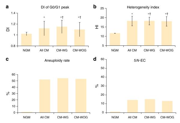
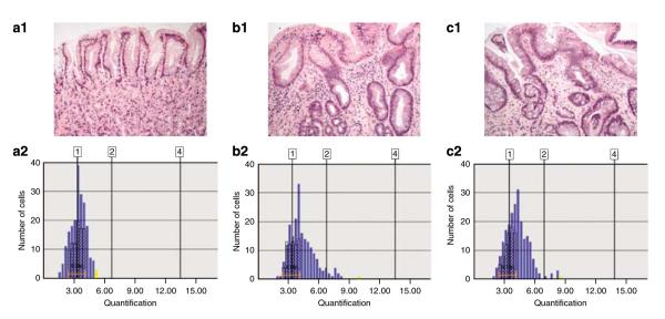
Methods: Mucosal biopsies of the esophagus from 68 patients with columnar metaplasia of the esophagus (22 without goblet cells and 46 with goblet cells) and 19 patients with normal gastric mucosa (controls) were histologically evaluated for the density of goblet cells. The latter group was divided into low-density, high-density, and very high-density goblet cell subgroups. Tissue sections of non-goblet epithelium and goblet cell epithelium (where present) were evaluated by image cytometry, and high-fidelity DNA histograms were created to indicate the G0/G1 peak DNA index (DI), DNA content heterogeneity index (HI), and the percentage of cells with DNA exceeding 5N (5N-EC). G0/G1 peaks with DI>1.1 were considered aneuploid.
Results: Normal gastric controls showed a mean peak DI of 1.02+/-0.03 and an HI of 11.6+/-0.7. None of the controls revealed aneuploidy or 5N-EC. Patients with metaplastic columnar epithelium with goblet cells showed a DI of 1.15+/-0.12, HI of 18.2+/-2.1, mild aneuploidy in 54% of the cases, and 5N-EC in 15% of the cases, all of which were significantly higher than in controls. Patients with metaplastic columnar epithelium without goblet cells showed DNA content results statistically similar to those of patients with metaplastic columnar epithelium with goblet cells, and also revealed significantly higher values compared with those of controls. Furthermore, there were no significant differences in any of the key DNA content abnormalities between non-goblet and goblet cell-containing epithelium in patients with metaplastic columnar epithelium with goblet cells, or between these two types of epithelium according to the density of goblet cells.
Conclusions: DNA content abnormalities occur with equal frequency and extent in metaplastic columnar epithelium of the esophagus without goblet cells compared with metaplastic columnar epithelium with goblet cells. These findings suggest that metaplastic non-goblet columnar epithelium of the esophagus may have neoplastic potential.
Figures
Comment in
-
Editorial: Defining a bad Barrett's segment: is it dependent on goblet cells?Am J Gastroenterol. 2009 Apr;104(4):825-7. doi: 10.1038/ajg.2009.90. Epub 2009 Mar 17. Am J Gastroenterol. 2009. PMID: 19343024
-
[Barrett's without goblet cells: a finding with clinical relevance?!].Z Gastroenterol. 2009 Aug;47(8):773-4. doi: 10.1055/s-0028-1109623. Epub 2009 Aug 6. Z Gastroenterol. 2009. PMID: 19662591 German. No abstract available.
-
Barrett's: do we still need goblet cells for diagnosis?Am J Gastroenterol. 2009 Sep;104(9):2355-6; author reply 2356-7. doi: 10.1038/ajg.2009.364. Am J Gastroenterol. 2009. PMID: 19727095 No abstract available.
References
-
- Hornick JL, Odze RD. Neoplastic precursor lesions in Barrett’s esophagus. Gastroenterol Clin North Am. 2007;36:775–96. - PubMed
-
- Yousef F, Cardwell C, Cantwell MM, et al. The incidence of esophageal cancer and high-grade dysplasia in Barrett’s esophagus: a systematic review and meta-analysis. Am J Epidemiol. 2008;168:237–49. - PubMed
-
- Wang KK, Sampliner RE. Practice Parameters Committee of the American College of Gastroenterology. Updated guidelines 2008 for the diagnosis, surveillance and therapy of Barrett’s esophagus. Am J Gastroenterol. 2008;103:788–97. - PubMed
-
- Sharma P, McQuaid K, Dent J, et al. A critical review of the diagnosis and management of Barrett’s esophagus: the AGA Chicago workshop. Gastro enterology. 2004;127:310–30. - PubMed
Publication types
MeSH terms
Substances
Grants and funding
LinkOut - more resources
Full Text Sources
