. 2009;4(3):e4848.
doi: 10.1371/journal.pone.0004848. Epub 2009 Mar 18.

Detection of CWD prions in urine and saliva of deer by transgenic mouse bioassay

Affiliations

Detection of CWD prions in urine and saliva of deer by transgenic mouse bioassay

Nicholas J Haley et al. PLoS One. 2009.

Abstract

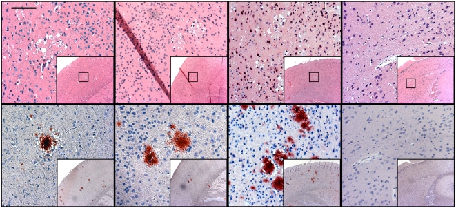
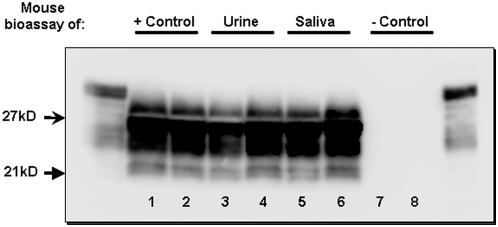
Chronic wasting disease (CWD) is a prion disease affecting captive and free-ranging cervids (e.g. deer, elk, and moose). The mechanisms of CWD transmission are poorly understood, though bodily fluids are thought to play an important role. Here we report the presence of infectious prions in the urine and saliva of deer with chronic wasting disease (CWD). Prion infectivity was detected by bioassay of concentrated, dialyzed urine and saliva in transgenic mice expressing the cervid PrP gene (Tg[CerPrP] mice). In addition, PrP(CWD) was detected in pooled and concentrated urine by protein misfolding cyclic amplification (PMCA). The concentration of abnormal prion protein in bodily fluids was very low, as indicated by: undetectable PrP(CWD) levels by traditional assays (western blot, ELISA) and prolonged incubation periods and incomplete TSE attack rates in inoculated Tg(CerPrP) mice (373(+/-)3 days in 2 of 9 urine-inoculated mice and 342(+/-)109 days in 8 of 9 saliva-inoculated mice). These findings help extend our understanding of CWD prion shedding and transmission and portend the detection of infectious prions in body fluids in other prion infections.


Conflict of interest statement

Competing Interests: The authors have declared that no competing interests exist.

Figures

Figure 1
Figure 1. Spongiform degeneration and PrPCWD identified by histopathology and immunohistochemistry.
Vacuolated neurons and spongiform degeneration of the neuropil characteristic of a TSE is evident on H&E staining, with the colocalization of PrPCWD specific immunostaining of florid plaques in the cortices of mice inoculated with positive control inoculum and concentrated urine and saliva from CWD-infected cervids. Negative control mice showed no evidence of spongiform degeneration or PrPCWD immunostaining. HRP-conjugated BAR-224 was used as a primary antibody. (Measure bar, 50 µm).
Figure 2
Figure 2. Western Blot detection of PrPCWD in urine and saliva-inoculated mice.
Western blotting analysis of control and test mice, demonstrating PrPCWD in positive control mice (lanes 1 and 2), as well as urine (lanes 3 and 4) and saliva (lanes 5 and 6) inoculated mice. Protease-resistant prions were not detected in negative control mice (lanes 7 and 8). Flanking lanes represent undigested PrPC.
Figure 3
Figure 3. Histopathologic evaluation of renal tissues from donor cervids.
(A) Minimal, chronic and proliferative glomerular disease and (B) mild interstitial fibrosis and lymphocytic infiltration were observed in 4 out of 5 donor deer. The remaining deer showed evidence of mild lymphocytic glomerulonephritis (C) as well as “tubular proteinosis.” (D, arrows).
Figure 4
Figure 4. Serial PMCA amplification of PrPCWD in concentrated deer urine and in the brains of urine-inoculated mice.
A) PrPCWD was detectable by serial PMCA (sPMCA) in control and urine inocula (lanes 1 and 2, respectively), while PrPCWD could not be identified in saliva and negative control inocula (lanes 3 and 4, respectively) after 3 rounds of amplification. B) Three rounds of sPMCA also amplified PrPCWD in the brains of CWD-infected mice, including positive-control inoculated mice and a single mouse inoculated with lyophilized urine (lanes 1 and 3, respectively). PrPCWD was not amplified in mice inoculated with negative control material (lanes 5 and 6) or in other mice inoculated with either urine (lane 2) or saliva (lane 4) from CWD+ deer. All flanking lanes represent undigested PrPC.

References

    1. Williams ES, Young S. Chronic wasting disease of captive mule deer: a spongiform encephalopathy. J Wildl Dis. 1980;16:89–98. - PubMed
    1. Williams ES, Young S. Spongiform encephalopathy of Rocky Mountain elk. J Wildl Dis. 1982;18:465–471. - PubMed
    1. Sigurdson CJ. A prion disease of cervids: chronic wasting disease. Vet Res. 2008;39:41. - PubMed
    1. Williams ES. Chronic wasting disease. Vet Pathol. 2005;42:530–549. - PubMed
    1. Miller MW, Williams ES, Hobbs NT, Wolfe LL. Environmental sources of prion transmission in mule deer. Emerg Infect Dis. 2004;10:1003–1006. - PMC - PubMed

Publication types