. 2009;2(5):433-43.
Epub 2009 Jan 30.

HIV-1 Tat contributes to Alzheimer's disease-like pathology in PSAPP mice

Affiliations

HIV-1 Tat contributes to Alzheimer's disease-like pathology in PSAPP mice

Brian Giunta et al. Int J Clin Exp Pathol. 2009.

Abstract

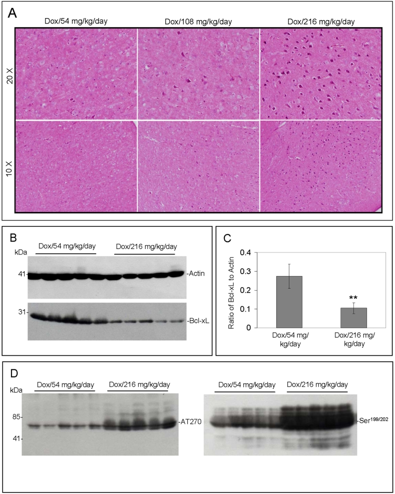
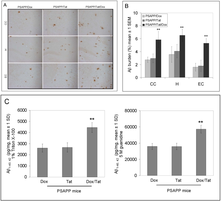
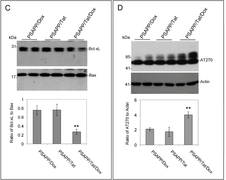
Prevalence of HIV-associated cognitive impairment is rising. Amyloid-beta (A-beta) plaque deposition in the brain may be a contributing factor as epidemiological data suggests significant numbers of long-term HIV survivors are at elevated risk of developing Alzheimer's disease (AD). HIV-1 Tat-induced A-beta deposition, tau phosphorylation, and subsequent neuronal death could be risk factors for subsequent AD and/or HIV-related cognitive impairment. To mimic this clinical condition, we generated mice with HIV-1 Tat-induced AD-like pathology. We first performed a short-term Doxycycline (dox) dosing (54, 108, and 216 mg/kg/day) study in transgenic mice whose astrocytes express HIV-1 Tat via activation of a GFAP/dox-inducible promoter. After one week, mouse brains were examined histologically and the expression of Bcl-xL, Bax, and phospho-tau was investigated by Western blotting. We next cross-bred these mice with the PSAPP mouse model of AD. To simulate chronic Tat secretion over periods longer than one week, we used an optimized dose of 54 mg/kg/day on a biweekly basis over three months; based on the initial dose ranging study in the Tat transgenic mice. This was followed by antisera detection of A-beta, and Western blot for phospho-tau, Bcl-xL, and Bax. Tat significantly induced neuron degeneration and tau phosphorylation in Tat transgenic mice, dox dependently (P<0.001) with the most robust effects at the 216 mg/kg/day dose. In the long term study, similar effects at the chronic 54 mg/kg/day dose were observed in PSAPP/Tat mice induced with dox. These mice also showed significantly more A-beta deposition (P < 0.05), neurodegeneration, neuronal apoptotic signaling, and phospho-tau than PSAPP mice (P < 0.05). In conclusion, HIV-1 Tat significantly promotes AD-like pathology in PSAPP/Tat mice. This model may provide a framework in which to identify new mechanisms involved in cognitive impairment in the HIV infected population, and possible treatments. Additional works will be needed to fully characterize the mechanism(s) of HIV- induced amyloid deposition, and also to uncover viral mechanisms promoting AD-like pathology in general.

Keywords: Alzheimer's; Dementia; HIV-1; PSAPP; Tat; beta-amyloid.


Figures

Figure 1
Figure 1
Oral administration of dox results in increased neuronal damage and tau phosphorylation with decreased Bcl-xL expression dose dependently in GT-tg mice. A. Mouse brain coronal frozen sections were stained with H&E. Left column indicates PSAPP mice receiving dox, middle column indicates PSAPP/Tat mice without dox and right column indicates PSAPP/Tat mice receiving dox. B. Bcl-xL expression was analyzed by Western blotting analysis of the mouse brain homogenates with anti-Bcl-xL antibody. C. Densitometry analysis shows the band density ratio of Bcl-xL to actin. D. Western blot analysis by antibody AT270 (left) or Ser199/202 (right) shows phosphorylated tau protein.
Figure 2
Figure 2
PSAPP/Tat mice receiving oral dox show increased A-beta plaques compared to PSAPP mice. A. Mouse brain coronal frozen sections were stained with rabbit polyclonal anti-human A antibody. Left column indicates PSAPP mice receiving dox, middle column indicates PSAPP/Tat mice without dox and right column indicates PSAPP/Tat mice receiving dox. As indicated, the top panels are from the cingulate cortex (CC), the middle panels are from the hippocampus (H), and bottom panels are from the entorhinal cortex (EC). B. Percentages of A antibody-immunoreactive A plaque (mean ± SD) were calculated by quantitative image analysis. C. Soluble A1-40, 42 (left) and insoluble A-beta1-40, 42 (right) prepared with 5 M guanidine were analyzed by ELISA. Data are presented as (pg/mg protein) of A1-40 or A1-42 separately.
Figure 3
Figure 3
PSAPP/Tat mice receiving oral dox show increased neuronal degeneration and tau-phosphorylation after oral administration of dox compared to PSAPP mice. Left column indicates PSAPP mice receiving dox, middle column indicates PSAPP/Tat mice without dox and right column indicates PSAPP/Tat mice receiving dox. Mouse brain coronal frozen sections were stained with H&E (A) or with phospho-tau antibody (AT270) (B). PSAPP/Tat mice receiving oral dox show increased neuronal degeneration and tau-phosphorylation after oral administration of dox compared to PSAPP mice. Mouse brain homogenates were prepared from these mice and subjected to Western blot analysis for Bcl-xL, Bax; and phosphor-Tau. Compared to PSAPP mice receiving dox or PSAPP/Tat mice without dox, PSAPP/Tat mice receiving dox demonstrate a significantly increased ratio of Bax to Bcl-xL (C) as well as Tau protein (phospho-AT270) to actin ratio by densitometry analyses (D).
Figure 3
Figure 3
PSAPP/Tat mice receiving oral dox show increased neuronal degeneration and tau-phosphorylation after oral administration of dox compared to PSAPP mice. Left column indicates PSAPP mice receiving dox, middle column indicates PSAPP/Tat mice without dox and right column indicates PSAPP/Tat mice receiving dox. Mouse brain coronal frozen sections were stained with H&E (A) or with phospho-tau antibody (AT270) (B). PSAPP/Tat mice receiving oral dox show increased neuronal degeneration and tau-phosphorylation after oral administration of dox compared to PSAPP mice. Mouse brain homogenates were prepared from these mice and subjected to Western blot analysis for Bcl-xL, Bax; and phosphor-Tau. Compared to PSAPP mice receiving dox or PSAPP/Tat mice without dox, PSAPP/Tat mice receiving dox demonstrate a significantly increased ratio of Bax to Bcl-xL (C) as well as Tau protein (phospho-AT270) to actin ratio by densitometry analyses (D).

Similar articles

Cited by

References

    1. Valcour V, Paul R. HIV infection and dementia in older adults. Clin Infect Dis. 2006;42:1449–1454. - PubMed
    1. Levy-Dweck S. HIV/AIDS fifty and older: A hidden and growing population. J Gerontol Soc Work. 2005;46:37–50. - PubMed
    1. Stoff DM., Khalsa JH, Monjan A, Portegies P. Introduction: HIV/AIDS and aging. AIDS. 2004;18(Suppl 1):S1–2. - PubMed
    1. Green DA, Masliah E, Vinters HV, Beizai P, Moore DJ, Achim CL. Brain deposition of beta-amyloid is a common pathologic feature in HIV positive patients. AIDS. 2005;19:407–411. - PubMed
    1. Achim C, Masliah E, Vinters H, Schindelar J, Green D. Society of Neuroscience. Washington, DC: 2004. Beta-amyloid in the HIV brain.

LinkOut - more resources