A comparison of angled sagittal MRI and conventional MRI in the diagnosis of herniated disc and stenosis in the cervical foramen
- PMID: 19294432
- PMCID: PMC2899504
- DOI: 10.1007/s00586-009-0932-x
A comparison of angled sagittal MRI and conventional MRI in the diagnosis of herniated disc and stenosis in the cervical foramen
Abstract
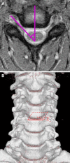
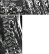
The object of this study is to demonstrate that angled sagittal magnetic resonance imaging (MRI) enables the precise diagnosis of herniated disc and stenosis in the cervical foramen, which is not available with conventional MRI. Due to both the anatomic features of the cervical foramen and the limitations of conventional MR techniques, it has been difficult to identify disease in the lateral aspects of the spinal canal and foramen using only conventional MRI. Angled sagittal MRI oriented perpendicular to the true course of the foramina facilitates the identification of the lateral disease. A review of 43 patients, who underwent anterior cervical discectomy and interbody fusion, is presented with a herniated disc and/or stenosis in the cervical foramen. They all had undergone conventional MRI and angled sagittal MRI. Fifty levels were surgically explored for evidence of foraminal herniated disc and stenosis. The results of each test were correlated with what was found at each explored surgical level. The sensitivity, specificity, and accuracy of both examinations for making the diagnosis of foraminal herniated disc and stenosis were compared. During the diagnosis of foraminal herniated disc, the sensitivity, specificity, and accuracy of angled sagittal MRI were 96.7, 95.0, and 96.0%, respectively, compared with 56.7, 85.0, and 68.0% for conventional MRI. In making the diagnosis of foraminal stenosis, the sensitivity, specificity, and accuracy of angled sagittal MRI were 96.3, 95.7, and 96.0%, respectively, compared with 40.7, 91.3, and 66.0% for conventional MRI. In the above groups, the difference between the tests for making the diagnosis of both foraminal herniated disc and stenosis was found to be statistically significant in sensitivity and accuracy. Angled sagittal MRI was a more accurate test compared to conventional MRI for making the diagnosis of herniated disc and stenosis in the cervical foramen. It can be utilized for the precise diagnosis of foraminal herniated disc and stenosis difficult or ambiguous in conventional MRI.
Figures


References
-
- Bischoff RJ, Rodriguez RP, Gupta K, Righi A, Dalton JE, Whitecloud TS. A comparison of computed tomography-myelography, magnetic resonance imaging, and myelography in the diagnosis of herniated nucleus pulposus and spinal stenosis. J Spinal Disord. 1993;6:289–295. doi: 10.1097/00002517-199306040-00002. - DOI - PubMed
-
- Edelman RR, Stark DD, Saini S, Ferrucci JT, Jr, Dinsmore RE, Ladd W, et al. Oblique planes of section in MR imaging. Radiology. 1986;159:807–810. - PubMed
Publication types
MeSH terms
LinkOut - more resources
Full Text Sources
Medical

