. 2009 Dec;5(4):447-60.
doi: 10.1007/s11302-009-9147-1. Epub 2009 Mar 18.

ATP, P2 receptors and the renal microcirculation

Affiliations

ATP, P2 receptors and the renal microcirculation

Edward W Inscho. Purinergic Signal. 2009 Dec.

Abstract

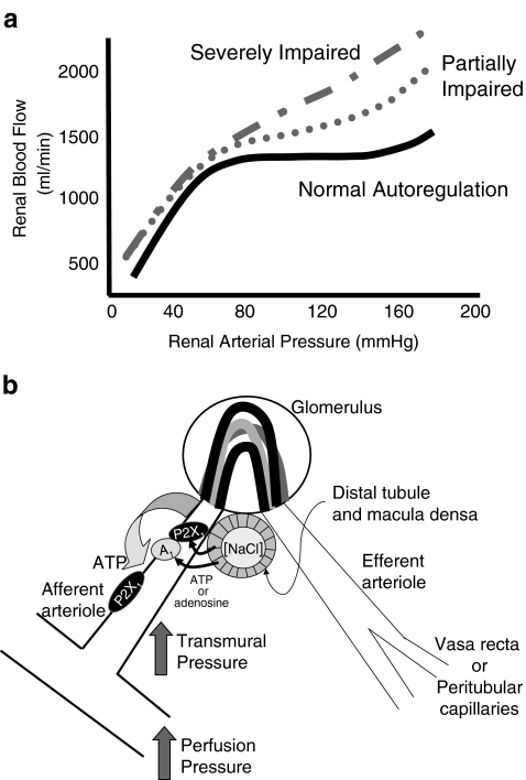
Purinoceptors are rapidly becoming recognised as important regulators of tissue and organ function. Renal expression of P2 receptors is broad and diverse, as reflected by the fact that P2 receptors have been identified in virtually every major tubular/vascular element. While P2 receptor expression by these renal structures is recognised, the physiological functions that they serve remains to be clarified. Renal vascular P2 receptor expression is complex and poorly understood. Evidence suggests that different complements of P2 receptors are expressed by individual renal vascular segments. This unique distribution has given rise to the postulate that P2 receptors are important for renal vascular function, including regulation of preglomerular resistance and autoregulatory behaviour. More recent studies have also uncovered evidence that hypertension reduces renal vascular reactivity to P2 receptor stimulation in concert with compromised autoregulatory capability. This review will consolidate findings related to the role of P2 receptors in regulating renal microvascular function and will present areas of controversy related to the respective roles of ATP and adenosine in autoregulatory resistance adjustments.


Figures

Fig. 1
Fig. 1
ATP concentration–response relationship for the intrarenal pre- and postglomerular vascular segments. Average segmental diameter responses evoked by ATP applied to the adventitial surface of arcuate arteries (top panel), interlobular arteries (second panel), afferent arterioles (third panel) and efferent arterioles (bottom panel). After the control period (Con), increasing concentrations of ATP (0.1, 1.0, 10 and 100 µM) were applied at 5-min intervals as indicated by the dotted lines. Each protocol ended with a 5-min recovery period (Rec) while bathed with control solution. Each data point represents diameter measurements taken at 12-s intervals and normalised as a percentage of the control diameter. Data were modified from an earlier report [21]
Fig. 2
Fig. 2
The effect of α β-methylene ATP and ATP on intracellular calcium concentration in preglomerular smooth muscle cells. Response of intracellular calcium concentration evoked by α β-methylene ATP or ATP (each 10 µM) in the presence of 1.8 mM extracellular calcium (a and b), in the absence of extracellular calcium (c and d) and during P2X1 receptor blockade with NF-279 (e). The periods of exposure to α β-methylene ATP or ATP administration are indicated by the black bars. The periods of exposure to calcium-free medium or NF-279 are shown by a second set of black bars. Calcium measurements were performed using fura 2. Data were modified from an earlier report [74]
Fig. 3
Fig. 3
Postulated signalling mechanisms for ATP-mediated autoregulatory adjustments in afferent arteriolar diameter. Panel a illustrates a normal profile for autoregulation of renal blood flow (solid black line) and the autoregulatory profiles that might be observed under conditions of moderate and severe autoregulatory dysfunction (modified from [171]). b ATP might be released from afferent arteriole smooth muscle cells in response to an increase in renal perfusion pressure. This ATP released into the perivascular interstitial fluid could act upon P2X1 receptors to evoke autoregulatory vasoconstrictor responses. ATP released from the macula densa, in response to an increase in distal tubular fluid NaCl concentration, can traverse the interstitial fluid of the juxtaglomerular apparatus to act on afferent arterioles and induce tubuloglomerular feedback-mediated vasoconstriction. Alternatively, released ATP could be degraded to adenosine in the interstitial fluid prior to activating afferent arteriolar adenosine A1 receptors to regulate afferent arteriolar resistance
Fig. 4
Fig. 4
Effect of P2 receptor blockade on the afferent arteriolar autoregulatory response induced by an increase in perfusion pressure. Pressure-mediated afferent arteriolar autoregulatory responses are depicted before and during P2 receptor blockade with NF-279 (top panel), PPADS (centre panel) and suramin (bottom panel). Autoregulatory responses were induced by increasing perfusion pressure in 30 mmHg increments at 5-min intervals. The data are expressed as a percentage of the control diameter and each data point represents the average diameter response over the last 2 min of each period. Control responses are shown by the black symbols and the response during P2 receptor blockade are shown by the grey symbols. At the end of each protocol, perfusion pressure was returned to 100 mmHg to determine recovery for the pressure stimulus. Data are modified from earlier reports [33, 51]
Fig. 5
Fig. 5
Effect of ANG-II hypertension on the afferent arteriolar diameter and calcium signalling responses to ATP. Panel a presents the changes in afferent arteriolar diameter in kidneys from normotensive rats (black symbols) and from Ang-II hypertensive rats (grey symbols). Data are expressed as a percentage of the control diameter. ATP was administered in concentrations of 0.1–100 μM. Values represent average diameters measured over the last 2 min of each treatment period and plotted as means ± SE. *P < 0.05, significant difference from control diameter; #P < 0.05 significant difference from normotensive controls. b Presents the change in intracellular calcium concentration (detected by fura 2) induced by 10 µM ATP in single preglomerular smooth muscle cells taken from normotensive (black trace) and ANG II hypertensive rats (grey trace). The period of ATP administration is depicted by the black bar. Data are modified from an earlier report [11]

Similar articles

Cited by

References

    1. {'text': '', 'ref_index': 1, 'ids': [{'type': 'PubMed', 'value': '10869730', 'is_inner': True, 'url': 'https://pubmed.ncbi.nlm.nih.gov/10869730/'}]}
    2. Bailey MA, Hillman KA, Unwin RJ (2000) P2 receptors in the kidney. J Auton Nerv Syst 81:264–270 - PubMed
    1. {'text': '', 'ref_index': 1, 'ids': [{'type': 'PubMed', 'value': '11044209', 'is_inner': True, 'url': 'https://pubmed.ncbi.nlm.nih.gov/11044209/'}]}
    2. Bailey MA, Imbert-Teboul M, Turner C, Marsy S, Srai K, Burnstock G, Unwin RJ (2000) Axial distribution and characterization of basolateral P2Y receptors along the rat renal tubule. Kidney Int 58:1893–1901 - PubMed
    1. {'text': '', 'ref_index': 1, 'ids': [{'type': 'PubMed', 'value': '15056981', 'is_inner': True, 'url': 'https://pubmed.ncbi.nlm.nih.gov/15056981/'}]}
    2. Bailey MA, Turner CM, Hus-Citharel A, Marchetti J, Imbert-Teboul M, Milner P, Burnstock G, Unwin R (2004) P2Y receptors present in the native and isolated rat glomerulus. Nephron Physiol 96:79–90 - PubMed
    1. {'text': '', 'ref_index': 1, 'ids': [{'type': 'PubMed', 'value': '9575906', 'is_inner': True, 'url': 'https://pubmed.ncbi.nlm.nih.gov/9575906/'}]}
    2. Chan CM, Unwin RJ, Bardini M, Oglesby IB, Ford APDW, Townsend-Nicholson A, Burnstock G (1998) Localization of the P2X1 purinoceptors by autoradiography and immunohistochemistry in the rat kidney. Am J Physiol Renal Physiol 274:F799–F804 - PubMed
    1. {'text': '', 'ref_index': 1, 'ids': [{'type': 'PubMed', 'value': '9639035', 'is_inner': True, 'url': 'https://pubmed.ncbi.nlm.nih.gov/9639035/'}]}
    2. Chan CM, Unwin RJ, Burnstock G (1998) Potential functional roles of extracellular ATP in kidney and urinary tract. Exp Nephrol 6:200–207 - PubMed

LinkOut - more resources