

Review
doi: 10.3748/wjg.15.1301.
Diagnosis of hepatocellular carcinoma
Affiliations
- PMID: 19294759
- PMCID: PMC2658831
- DOI: 10.3748/wjg.15.1301
Item in Clipboard
Review
Diagnosis of hepatocellular carcinoma
World J Gastroenterol.
.
Abstract
Hepatocellular carcinoma (HCC) is one of the commonest cancers worldwide, particularly in parts of the developing world, and is increasing in incidence. This article reviews the current modalities employed for the diagnosis of HCC, including serum markers, radiological techniques and histological evaluation, and summarises international guidelines for the diagnostic approach to HCC.
Figures
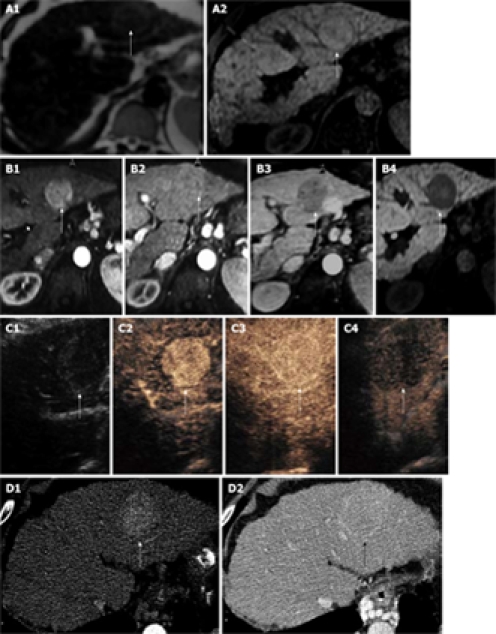
There is a large HCC in the left lobe of the liver with a pseudocapsule, hyper-enhancing on arterial phase and showing washout on late phases on MR and CEUS and is iso-intense in portal phases on CT and CEUS. The pseudo-capsule enhances in the portal phase on all modalities. A1: T2 weighted scan showing slightly higher intensity HCC (arrow); A2: T1 weightted scan shows same HCC which is iso-intense (arrow); B1: MultiHance enhanced T1 weighted scan in the arterial phase showing enhancement of the HCC (arrow); B2: MultiHance enhanced T1 weighted scan in the portal phase showing iso-enhancement of the HCC (arrow); B3: MultiHance enhanced T1 weighted scan at 2 min showing contrast wash-out in the HCC (arrow); B4: MultiHance enhanced T1 weighted scan at 40 min showing hypoiintense HCC (arrow); C1: Baseline ultrasound shows iso-echoic HCC (arrow); C2: SonoVue enhanced ultrasound shows hyper-enhancing HCC (arrow) in the arterial phase; C3: SonoVue enhanced ultrasound shows iso-enhancing HCC (arrow) in the portal phase with enhancement of the pseudocapsule; C4: SonoVue enhanced ultrasound shows wash-out of the HCC (arrow) in the late phase; D1: Contrast-enhanced CT scan shows enhancement of the HCC (arrow) in the arterial phase; D2: Contrast-enhanced CT scan shows iso-enhancement of the HCC (arrow) in the portal phase with enhancement of the pseudocapsule.
Similar articles
-
Evaluation of squamous cell carcinoma antigen-immunoglobulin M complex (SCCA-IGM) and alpha-L-fucosidase (AFU) as novel diagnostic biomarkers for hepatocellular carcinoma.Tumour Biol. 2014 Nov;35(11):11559-64. doi: 10.1007/s13277-014-2467-y. Epub 2014 Aug 17. Tumour Biol. 2014. PMID: 25129443
-
Screening for hepatocellular carcinoma.Eur J Gastroenterol Hepatol. 1996 Sep;8(9):856-60. Eur J Gastroenterol Hepatol. 1996. PMID: 8889450 Review.
-
VWF/ADAMTS13 ratio as a potential biomarker for early detection of hepatocellular carcinoma.BMC Gastroenterol. 2019 Oct 21;19(1):167. doi: 10.1186/s12876-019-1082-1. BMC Gastroenterol. 2019. PMID: 31638892 Free PMC article.
-
Circulating predictive and diagnostic biomarkers for hepatitis B virus-associated hepatocellular carcinoma.World J Gastroenterol. 2016 Oct 7;22(37):8271-8282. doi: 10.3748/wjg.v22.i37.8271. World J Gastroenterol. 2016. PMID: 27729734 Free PMC article. Review.
-
Clinical biomarkers in hepatocellular carcinoma (HCC).Front Biosci (Schol Ed). 2010 Jan 1;2(2):571-7. doi: 10.2741/s86. Front Biosci (Schol Ed). 2010. PMID: 20036969 Review.
Cited by
-
Glycine-Poly-L-Lactic Acid Copolymeric Nanoparticles for the Efficient Delivery of Bortezomib.Pharm Res. 2019 Sep 13;36(11):160. doi: 10.1007/s11095-019-2686-4. Pharm Res. 2019. PMID: 31520196
-
Controlling liver cancer internationally: A qualitative study of clinicians' perceptions of current public policy needs.Health Res Policy Syst. 2011 Jul 28;9:32. doi: 10.1186/1478-4505-9-32. Health Res Policy Syst. 2011. PMID: 21798002 Free PMC article.
-
Hepatocellular Carcinoma: Tumorigenesis and Prediction Markers.Gastroenterology Res. 2009 Aug;2(4):191-199. doi: 10.4021/gr2009.07.1304. Epub 2009 Jul 20. Gastroenterology Res. 2009. PMID: 27942274 Free PMC article. Review.
-
Quantitative mass spectrometric analysis of glycoproteins combined with enrichment methods.Mass Spectrom Rev. 2015 Mar-Apr;34(2):148-65. doi: 10.1002/mas.21428. Epub 2014 Jun 2. Mass Spectrom Rev. 2015. PMID: 24889823 Free PMC article. Review.
-
Diagnostic Value of Circulating microRNAs for Hepatocellular Carcinoma: Results of a Meta-analysis and Validation.Biochem Genet. 2025 Jan 3. doi: 10.1007/s10528-024-11001-2. Online ahead of print. Biochem Genet. 2025. PMID: 39751721
References
-
- World Health Organization. Mortality database. Available from: http://www.who.int/whosis/en/
-
- Gupta S, Bent S, Kohlwes J. Test characteristics of alpha-fetoprotein for detecting hepatocellular carcinoma in patients with hepatitis C. A systematic review and critical analysis. Ann Intern Med. 2003;139:46–50. - PubMed
-
- França AV, Elias Junior J, Lima BL, Martinelli AL, Carrilho FJ. Diagnosis, staging and treatment of hepatocellular carcinoma. Braz J Med Biol Res. 2004;37:1689–1705. - PubMed
-
- Terentiev AA, Moldogazieva NT. Structural and functional mapping of alpha-fetoprotein. Biochemistry (Mosc) 2006;71:120–132. - PubMed
-
- Canick JA, MacRae AR. Second trimester serum markers. Semin Perinatol. 2005;29:203–208. - PubMed
Publication types
MeSH terms
Substances
Grants and funding
LinkOut - more resources
Full Text Sources
Other Literature Sources
Medical
Miscellaneous
