

Ex-vivo evaluation of gene therapy vectors in human pancreatic (cancer) tissue slices
- PMID: 19294766
- PMCID: PMC2658838
- DOI: 10.3748/wjg.15.1359
Ex-vivo evaluation of gene therapy vectors in human pancreatic (cancer) tissue slices
Abstract
Aim: To culture human pancreatic tissue obtained from small resection specimens as a pre-clinical model for examining virus-host interactions.
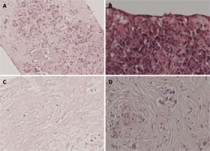
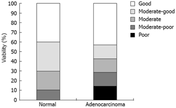
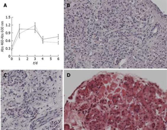
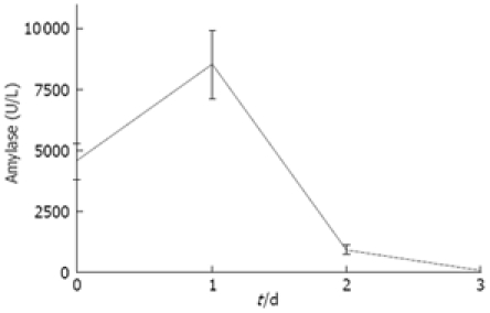
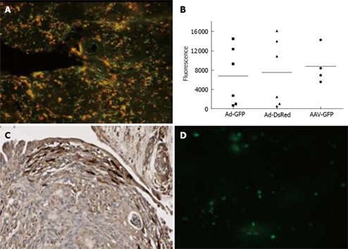
Methods: Human pancreatic tissue samples (malignant and normal) were obtained from surgical specimens and processed immediately to tissue slices. Tissue slices were cultured ex vivo for 1-6 d in an incubator using 95% O(2). Slices were subsequently analyzed for viability and morphology. In addition the slices were incubated with different viral vectors expressing the reporter genes GFP or DsRed. Expression of these reporter genes was measured at 72 h after infection.
Results: With the Krumdieck tissue slicer, uniform slices could be generated from pancreatic tissue but only upon embedding the tissue in 3% low melting agarose. Immunohistological examination showed the presence of all pancreatic cell types. Pancreatic normal and cancer tissue slices could be cultured for up to 6 d, while retaining viability and a moderate to good morphology. Reporter gene expression indicated that the slices could be infected and transduced efficiently by adenoviral vectors and by adeno associated viral vectors, whereas transduction with lentiviral vectors was limited. For the adenoviral vector, the transduction seemed limited to the peripheral layers of the explants.
Conclusion: The presented system allows reproducible processing of minimal amounts of pancreatic tissue into slices uniform in size, suitable for pre-clinical evaluation of gene therapy vectors.
Figures
Similar articles
-
Anti-viral state segregates two molecular phenotypes of pancreatic adenocarcinoma: potential relevance for adenoviral gene therapy.J Transl Med. 2010 Jan 29;8:10. doi: 10.1186/1479-5876-8-10. J Transl Med. 2010. PMID: 20113473 Free PMC article.
-
Specific targeting of pancreatic islet cells in vivo by insulin-promoter-driven adenoviral conjugated reporter genes.World J Surg. 2006 Aug;30(8):1543-52. doi: 10.1007/s00268-005-0688-3. World J Surg. 2006. PMID: 16855800
-
Initial Characterization of Integrase-Defective Lentiviral Vectors for Pancreatic Cancer Gene Therapy.Hum Gene Ther. 2016 Feb;27(2):184-92. doi: 10.1089/hum.2015.151. Hum Gene Ther. 2016. PMID: 26731312 Free PMC article.
-
Pancreatic carcinoma-specific immunotherapy using novel tumor specific cytotoxic T cells.Oncotarget. 2016 Dec 13;7(50):83601-83610. doi: 10.18632/oncotarget.13469. Oncotarget. 2016. PMID: 27876704 Free PMC article.
-
Gene therapy for pancreatic cancer targeting the genomic alterations of tumor suppressor genes using replication-selective oncolytic adenovirus.Hum Cell. 2002 Sep;15(3):138-50. doi: 10.1111/j.1749-0774.2002.tb00108.x. Hum Cell. 2002. PMID: 12703544 Review.
Cited by
-
Ex vivo functional whole organ in biomedical research: a review.J Artif Organs. 2025 Jun;28(2):131-145. doi: 10.1007/s10047-024-01478-4. Epub 2024 Nov 27. J Artif Organs. 2025. PMID: 39592544 Review.
-
Pancreas 3D Organoids: Current and Future Aspects as a Research Platform for Personalized Medicine in Pancreatic Cancer.Cell Mol Gastroenterol Hepatol. 2017 Dec 16;5(3):289-298. doi: 10.1016/j.jcmgh.2017.12.004. eCollection 2018 Mar. Cell Mol Gastroenterol Hepatol. 2017. PMID: 29541683 Free PMC article. Review.
-
Tumour-reprogrammed stromal BCAT1 fuels branched-chain ketoacid dependency in stromal-rich PDAC tumours.Nat Metab. 2020 Aug;2(8):775-792. doi: 10.1038/s42255-020-0226-5. Epub 2020 Jul 6. Nat Metab. 2020. PMID: 32694827 Free PMC article.
-
Long-lived pancreatic ductal adenocarcinoma slice cultures enable precise study of the immune microenvironment.Oncoimmunology. 2017 May 25;6(7):e1333210. doi: 10.1080/2162402X.2017.1333210. eCollection 2017. Oncoimmunology. 2017. PMID: 28811976 Free PMC article.
-
Hypoxia pathways and cellular stress activate pancreatic stellate cells: development of an organotypic culture model of thick slices of normal human pancreas.PLoS One. 2013 Sep 30;8(9):e76229. doi: 10.1371/journal.pone.0076229. eCollection 2013. PLoS One. 2013. PMID: 24098783 Free PMC article.
References
-
- Kuhlmann KF, de Castro SM, Wesseling JG, ten Kate FJ, Offerhaus GJ, Busch OR, van Gulik TM, Obertop H, Gouma DJ. Surgical treatment of pancreatic adenocarcinoma; actual survival and prognostic factors in 343 patients. Eur J Cancer. 2004;40:549–558. - PubMed
-
- Sultana A, Smith CT, Cunningham D, Starling N, Neoptolemos JP, Ghaneh P. Meta-analyses of chemotherapy for locally advanced and metastatic pancreatic cancer. J Clin Oncol. 2007;25:2607–2615. - PubMed
-
- Liu TC, Kirn D. Systemic efficacy with oncolytic virus therapeutics: clinical proof-of-concept and future directions. Cancer Res. 2007;67:429–432. - PubMed
-
- Heise C, Sampson-Johannes A, Williams A, McCormick F, Von Hoff DD, Kirn DH. ONYX-015, an E1B gene-attenuated adenovirus, causes tumor-specific cytolysis and antitumoral efficacy that can be augmented by standard chemotherapeutic agents. Nat Med. 1997;3:639–645. - PubMed
Publication types
MeSH terms
Substances
LinkOut - more resources
Full Text Sources
Medical
