. 2009 Mar;32(3):361-8.
doi: 10.1093/sleep/32.3.361.

Airway dilator muscle activity and lung volume during stable breathing in obstructive sleep apnea

Affiliations

Airway dilator muscle activity and lung volume during stable breathing in obstructive sleep apnea

Amy S Jordan et al. Sleep. 2009 Mar.

Abstract

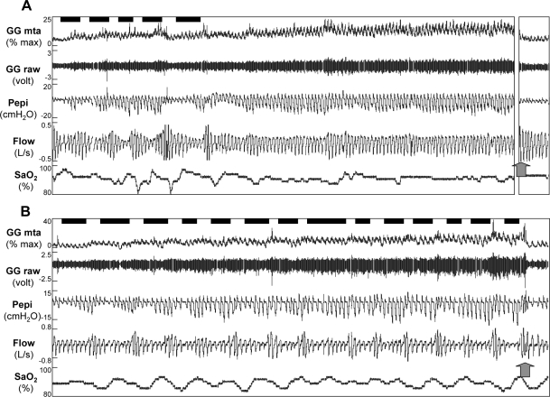
Study objectives: Many patients with obstructive sleep apnea (OSA) have spontaneous periods of stable flow limited breathing during sleep without respiratory events or arousals. In addition, OSA is often more severe during REM than NREM and more severe during stage 2 than slow wave sleep (SWS). The physiological mechanisms for these observations are unknown. Thus we aimed to determine whether the activity of two upper airway dilator muscles (genioglossus and tensor palatini) or end-expiratory lung volume (EELV) differ between (1) spontaneously occurring stable and cyclical breathing and (2) different sleep stages in OSA.

Design: Physiologic observation.

Setting: Sleep physiology laboratory.

Study participants: 15 OSA patients with documented periods of spontaneous stable breathing.

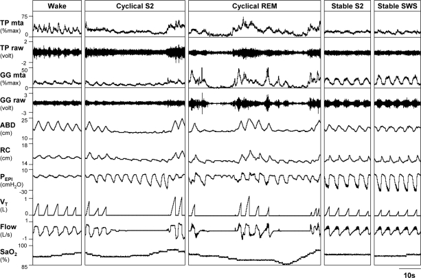
Intervention: Subjects were instrumented with intramuscular electrodes for genioglossus and tensor palatini electromyograms (EMG(GG) and EMG(TP)), chest and abdominal magnetometers (EELV measurement), an epiglottic pressure catheter (respiratory effort), and a mask and pneumotachograph (airflow/ventilation). Patients slept supine overnight without CPAP.

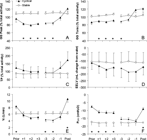
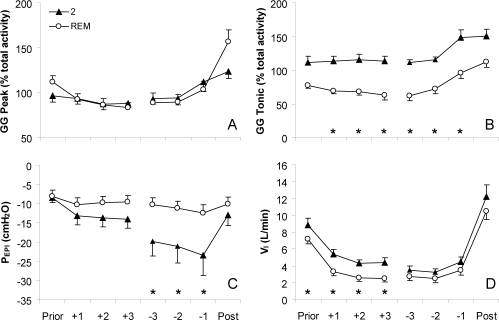
Measurements and results: Peak and Tonic EMG(GG) were significantly lower during cyclical (85.4 +/- 2.7 and 94.6 +/- 4.7 % total activity) than stable breathing (109.4 +/- 0.4 and 103 +/- 0.8% total activity, respectively). During respiratory events in REM, tonic EMG(GG) activity was lower than during respiratory events in stage 2 (71.9 +/- 5.1 and 119.6 +/- 5.6% total activity). EMG(GG) did not differ between stable stage 2 and stable SWS (98.9 +/- 3.2 versus 109.7 +/- 4.4% total activity), nor did EMG(TP) or EELV differ in any breathing condition/sleep stage.

Conclusions: Increased genioglossus muscle tone is associated with spontaneous periods of stable flow limited breathing in the OSA subjects studied. Reductions in genioglossus activity during REM may explain the higher severity of OSA in that stage. Increased lung volume and tensor palatini activity do not appear to be major mechanisms enabling spontaneous stable flow limited breathing periods.


Figures

Figure 1
Figure 1
Raw data from a 34 year old male patient with OSA (AHI = 57 events/h) during wakefulness, cyclical stage 2 (S2), cyclical REM, stable stage 2 (S2), and stable slow wave sleep (SWS). Note the elevated raw and moving time averaged (MTA) activity of the genioglossus (GG) but not tensor palatini (TP) during stable breathing, which is flow limited and accompanied by snoring (high frequency oscillation visible on Flow and PEPI during inspiration). Also note the unchanged rib cage (RC) and abdominal (ABD) positions and large negative epiglottic pressures (PEPI) generated. Airflow (Flow), tidal volume (VT), and arterial oxygen saturation (SpO2) indicate where respiratory events occur and the desaturation associated with the events.
Figure 2
Figure 2
Peak inspiratory (Peak) and expiratory tonic (Tonic) activity of the genioglossus (GG), tensor palatini (TP), end-expiratory lung volume (EELV), inspired minute ventilation (VI) and epiglottic pressure (PEPI) on the breath before (Prior) respiratory events, first (+1 to +3) and last (−3 to −1) 3 respiratory efforts during events and breath following (Post) respiratory events in stage 2 sleep and corresponding time periods (8 breath averages) of stable stage 2 sleep. Muscle activity is expressed as a percent of the average activity across both states in each subject (% total activity) due to high variability in the absolute level of muscle activity between subjects (range 3.9 to 38.2 % max). Mean (SEM) presented, n = 10. *indicates P < 0.05 between cyclical and stable conditions on these breaths/efforts by post hoc testing.
Figure 3
Figure 3
Peak inspiratory (Peak) and expiratory tonic (Tonic) activity of the genioglossus (GG), epiglottic pressure (PEPI) and inspired minute ventilation (VI) on the breath before (Prior) respiratory events, first (+1 to +3) and last ( −3 to −1) 3 respiratory efforts during events and breath following (Post) respiratory events in stage 2 and REM sleep. Muscle activity is expressed as a percent of the average activity across both states in each subject due to high variability in the absolute level of muscle activity between subjects (range 3.7 to 32.8 % max). Mean (SEM) presented, n = 12. *indicates P < 0.05 between sleep stages on these breaths/efforts by post hoc testing.
Figure 4
Figure 4
Raw data for 10 min after sleep onset in a 45-year-old man with OSA (AHI = 57.1 events/h) who developed stable breathing (A) and a 49 year old male patient (AHI = 69.7 events/h) who did not develop stable breathing (B). Arterial oxygen saturation (SpO2), airflow (Flow), epiglottic pressure (PEPI), and both the raw and moving time averaged (MTA) genioglossus (GG) electromyogram are shown. Respiratory events marked by the technician are shown by black bars above the GG signal. Note that both individuals had increased GG activity as sleep progressed. However, only subject A was able to stabilize breathing (although still flow limited). Following full awakening (as shown by the arrow), genioglossus activity instantly fell to baseline in both subjects. In Subject A, 6.5 min of data are omitted prior to full awakening (broken axis), such that 10 min of data are shown for both individuals.

References

    1. Young T, Blustein J, Finn L, Palta M. Sleep-disordered breathing and motor vehicle accidents in a population- based sample of employed adults. Sleep. 1997;20:608–13. - PubMed
    1. Peppard PE, Young T, Palta M, Skatrud J. Prospective study of the association between sleep-disordered breathing and hypertension. N Engl J Med. 2000;342:1378–84. - PubMed
    1. Shahar E, Whitney CW, Redline S, et al. Sleep-disordered breathing and cardiovascular disease: cross-sectional results of the Sleep Heart Health Study. Am J Respir Crit Care Med. 2001;163:19–25. - PubMed
    1. Stanchina ML, Malhotra A, Fogel RB, et al. Genioglossus muscle responsiveness to chemical and mechanical stimuli during non-rapid eye movement sleep. Am J Respir Crit Care Med. 2002;165:945–9. - PubMed
    1. Lo Y, Jordan AS, Malhotra A, et al. Genioglossal muscle response to CO2 stimulation during NREM sleep. Sleep. 2006;29:470–7. - PMC - PubMed

Publication types