. 2009 Apr 1;122(Pt 7):929-36.
doi: 10.1242/jcs.033928.

Laminin is required for Schwann cell morphogenesis

Affiliations

Laminin is required for Schwann cell morphogenesis

Wei-Ming Yu et al. J Cell Sci. .

Abstract

Development of the peripheral nervous system requires radial axonal sorting by Schwann cells (SCs). To accomplish sorting, SCs must both proliferate and undergo morphogenetic changes such as process extension. Signaling studies reveal pathways that control either proliferation or morphogenesis, and laminin is essential for SC proliferation. However, it is not clear whether laminin is also required for SC morphogenesis. By using a novel time-lapse live-cell-imaging technique, we demonstrated that laminins are required for SCs to form a bipolar shape as well as for process extension. These morphological deficits are accompanied by alterations in signaling pathways. Phosphorylation of Schwannomin at serine 518 and activation of Rho GTPase Cdc42 and Rac1 were all significantly decreased in SCs lacking laminins. Inhibiting Rac1 and/or Cdc42 activities in cultured SCs attenuated laminin-induced myelination, whereas forced activation of Rac1 and/or Cdc42 in vivo improved sorting and hypomyelinating phenotypes in SCs lacking laminins. These findings indicate that laminins play a pivotal role in regulating SC cytoskeletal signaling. Coupled with previous results demonstrating that laminin is critical for SC proliferation, this work identifies laminin signaling as a central regulator coordinating the processes of proliferation and morphogenesis in radial axonal sorting.


Figures
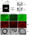
Fig. 1.
Fig. 1.
SCs lacking laminins do not ensheath or myelinate axons. (A) PCR analysis of genomic DNA of co-cultures from homozygous fLAM γ1 mice (F/F) infected with adenoviruses (Adv) expressing LacZ or Cre. The primers amplified the unrecombined (3.2 kb) and recombined (2.3 kb) fLAM γ1 alleles. (B) Myelination of mouse SC-DRG co-cultures infected with Ad-LacZ or Ad-Cre 8 days after addition of ascorbate or exogenous laminins was detected by immunostaining for laminins (Ln; green) and MBP (red) or by electron microscopy (EM). Scale bar: 50 μm in Ln/MBP, 1 μm in EM. (C) The expression of myelin protein zero (P0) in co-cultures 8 days (MF8) or 14 days (MF14) after addition of ascorbate was assessed by immunoblotting. β-Actin served as the loading control (con, control; mut, mutant; mut+Ln, mutant with laminins).
Fig. 2.
Fig. 2.
SCs lacking laminins fail to establish a bipolar morphology. Control (A), mutant (B), and mutant co-cultures with exogenous laminins (C) at MF8 were stained for neurofilment (NF) (red), S100 (green), and MBP (blue). Confocal microscopy was used, and the collected images were merged. (D) Comparison of SC length (measured by S100 staining) in co-cultures at MF8 (three fields per co-culture; six co-cultures in control and mutant+Ln; eight co-cultures in mutant; **P<0.001 compared with control). Broken lines represent the average. (E,F) Real-time analysis of the bipolar morphology of SCs. (E) Co-cultures were labeled with Ad-mCherry-NFL and Ad-eGFP-actin and imaged for 16 hours using a spinning disk confocal microscope. Five z-stacks were taken every 15 minutes. Each panel is a maximum projection of the z-stacks. (F) Quantification of the axial axis (parallel to axons) to radial axis (vertical to axons) ratio over a 15-hour period reveals that mutant SCs do not establish a bipolar morphology (duplicate assay; *P<0.05; error bars, s.e.m.). Scale bar: 20 μm in A-C, 17 μm in E.
Fig. 3.
Fig. 3.
SCs lacking laminins show decreased process extension. (A) SC-DRG co-cultures were labeled with Ad-mCherry-NFL and Ad-eGFP-actin and then imaged for 16 hours using a spinning disk confocal microscope. Ten z-stacks were taken every 5 minutes. Control SCs show extensive process extension (arrows), whereas mutants form fewer and shorter processes (arrowheads). Scale bar: 20 μm. (B,C) Quantification of process extension rate (B) and the length of processes (C) over a 12-hour period reveals that mutant SCs extend fewer (**P<0.001; error bars, s.e.m.) and shorter (**P<0.001; broken lines represent the average) processes.
Fig. 4.
Fig. 4.
SCs lacking laminins show aberrant cytoskeletal signaling. (A) Phosphorylation of Schwannomin at Ser518 in control (c), mutant (m), and mutant co-cultures with exogenous laminins (Ln) incubated for 1, 2 and 8 days in MF (MF1d, 2d and 8d, respectively) and in control and mutant sciatic nerves (SN) at postnatal day zero (P0) was assessed by immunoblot. (B) Quantitative analysis of phospho-Ser518 Schwannomin normalized with total Schwannomin level in co-cultures at MF2d and sciatic nerves. Triplicate assays; *P<0.05; error bars, s.e.m. (C) Activated Rac1 or Cdc42 of control (c), mutant (m), and mutant co-cultures with exogenous laminins (Ln) incubated for 12, 24 and 48 hours in MF (MF12hr, 24hr and 48hr, respectively) and in control and mutant sciatic nerves (SN) at P0 was assessed by affinity precipitation-immunoblot assay using PAK-1 PBD agarose. Total cell lysates were fractionated and probed with Rac1-Cdc42 antibody to detect total Rac1 and total Cdc42. (D) Quantitative analysis of affinity precipitation-immunoblot assay of co-cultures at MF12hr (for activated Rac1) or MF24hr (for activated Cdc42) and sciatic nerves shows that activated Rac1 and Cdc42 were both significantly higher in controls than in mutants. Addition of exogenous laminins to mutant co-cultures partially restored the levels of activated Rac1 and Cdc42. Signal intensity of activated Rac1 and Cdc42 (GTP-bound Rac1 and Cdc42) was measured by ImageJ and normalized to total Rac1 or total Cdc42 at the indicated time points. Triplicate assays; *P<0.05, **P<0.01; error bars, s.e.m.
Fig. 5.
Fig. 5.
SCs lacking laminins show decreased ErbB2 phosphorylation. (A) Phosphorylation of ErbB2 in control (c) and mutant (m) co-cultures before induction of myelination (MF0) and control (c), mutant (m), and mutant co-cultures with exogenous laminins (Ln) incubated for 1 or 2 days in MF (MF1d and MF2d, respectively) was assessed by immunoblot. (B) Quantitative analysis of phospho-ErbB2 normalized with total ErbB2 level in co-cultures at various time points. Triplicate assays; *P<0.05, **P<0.01; error bars, s.e.m. Phosphorylation of ErbB2 was decreased in co-cultures lacking laminins.
Fig. 6.
Fig. 6.
Expression of Rac1DN and/or Cdc42DN in SCs attenuates laminin-induced myelination. Mutant co-cultures with exogenous laminins infected with Ad-LacZ (A), Ad-Cdc42DN (B), Ad-Rac1DN (C), or Ad-Rac1DN and Ad-Cdc42DN (D) at MF14 were stained for MBP to detect myelin formation. Mutant co-cultures infected with Ad-LacZ (E) or Ad-Rac1CA and Ad-Cdc42CA (F) at MF14 were stained for MBP to detect myelin formation. Scale bar: 50 μm. (G,H) The expression of P0 in mutant co-cultures with (G) or without (H) exogenous laminins infected with various types of adenoviruses at MF14 was assessed by immunoblotting. β-Actin served as the loading control. (I) Quantitative analysis of immunoblot assay from (G) and (H). Signal intensity of P0 was measured by ImageJ and normalized to β-actin. Triplicate assays; **P<0.01, ***P<0.001 compared to signal intensity of mut+Ln. Myelination was decreased in mutant co-cultures treated with exogenous laminins that were infected with Ad-Cdc42DN and/or Ad-Rac1DN. Myelination was not significantly restored in mutant co-cultures infected with Ad-Rac1CA and Ad-Cdc42CA.
Fig. 7.
Fig. 7.
The myelinating and sorting phenotypes of SCs lacking laminins are improved after treatment of an adenovirus expressing Rac1CA and/or Cdc42CA. Adenoviruses expressing eGFP, Rac1CA and/or Cdc42CA were injected into the endoneurium of P8 mutant sciatic nerve. Validation of the treatment was confirmed by eGFP expression (B and C). eGFP was expressed in nerves treated with Ad-eGFP (C) but not in nerves injected with saline (B). Two weeks after treatment (P22), transverse semithin (D-G) or ultrathin (H-K) sections were examined 1.5 mm proximal to the injection site. In mutant nerves treated with Ad-Rac1CA, the average size of each unsorted axonal bundle was reduced as compared with Ad-eGFP-treated nerves (asterisks, E vs D). In mutant nerves treated with Cdc42CA or with both viruses (Rac1CA and Cdc42CA), the average size of each unsorted axonal bundle was reduced and the total number of sorted myelinated fibers was increased as compared with Ad-eGFP-treated nerves (F and G vs D). Electron micrographs of nerves treated with both Rac1CA and Cdc42CA demonstrate that newly sorted (shown by `a' in I) and myelinated fibers (arrows in K) were present when compared with Ad-eGFP-treated nerves (I and K vs H and J) (a, axon; B, unsorted bundle). Scale bar: 50 μm in B,C; 20 μm in D-G; 4 μm in H,I; 10 μmin J,K. (L) Statistical analysis revealed that the average area of each unsorted bundle significantly decreased in Rac1CA-treated nerves, Cdc42CA-treated nerves, and in nerves treated with Rac1CA and Cdc42CA (nine fields from three different animals in each group; *P<0.05, **P<0.01 by Student's t-test; error bars, s.e.m.). (M) Statistical analysis revealed that total sorted myelinated fibers significantly increased in Cdc42CA-treated nerves and in nerves treated with Rac1CA and Cdc42CA (nine fields from three different animals in each group; *P<0.05, **P<0.01 by Student's t-test; error bars, s.e.m.).

Similar articles

Cited by

References

    1. Benninger, Y., Thurnherr, T., Pereira, J. A., Krause, S., Wu, X., Chrostek-Grashoff, A., Herzog, D., Nave, K. A., Franklin, R. J., Meijer, D. et al. (2007). Essential and distinct roles for cdc42 and rac1 in the regulation of Schwann cell biology during peripheral nervous system development. J. Cell Biol. 177, 1051-1061. - PMC - PubMed
    1. Chen, Z. L. and Strickland, S. (2003). Laminin gamma1 is critical for Schwann cell differentiation, axon myelination, and regeneration in the peripheral nerve. J. Cell Biol. 163, 889-899. - PMC - PubMed
    1. Cosgaya, J. M., Chan, J. R. and Shooter, E. M. (2002). The neurotrophin receptor p75NTR as a positive modulator of myelination. Science 298, 1245-1248. - PubMed
    1. Feltri, M. L., Graus Porta, D., Previtali, S. C., Nodari, A., Migliavacca, B., Cassetti, A., Littlewood-Evans, A., Reichardt, L. F., Messing, A., Quattrini, A. et al. (2002). Conditional disruption of beta 1 integrin in Schwann cells impedes interactions with axons. J. Cell Biol. 156, 199-209. - PMC - PubMed
    1. Grove, M., Komiyama, N. H., Nave, K. A., Grant, S. G., Sherman, D. L. and Brophy, P. J. (2007). FAK is required for axonal sorting by Schwann cells. J. Cell Biol. 176, 277-282. - PMC - PubMed

Publication types

MeSH terms