. 2009 Apr 1;122(Pt 7):1025-34.
doi: 10.1242/jcs.041913.

Distinct role of PLCbeta3 in VEGF-mediated directional migration and vascular sprouting

Affiliations

Distinct role of PLCbeta3 in VEGF-mediated directional migration and vascular sprouting

Resham Bhattacharya et al. J Cell Sci. .

Abstract

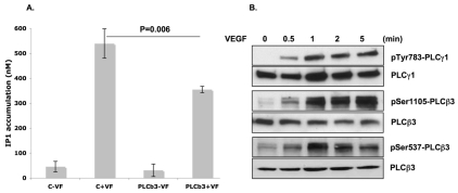
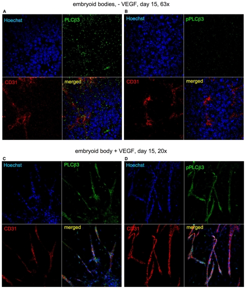
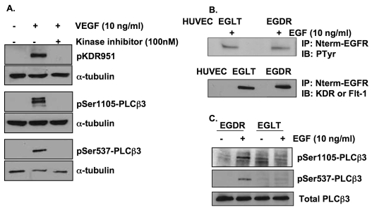
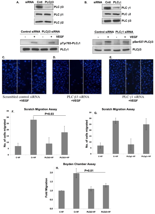
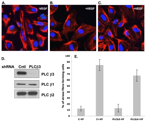
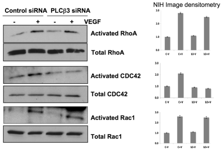
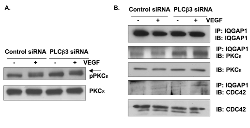
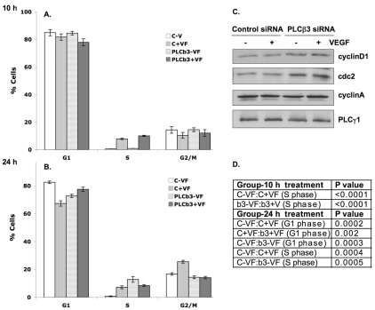
Endothelial cell proliferation and migration is essential to angiogenesis. Typically, proliferation and chemotaxis of endothelial cells is driven by growth factors such as vascular endothelial growth factor (VEGF) and basic fibroblast growth factor (bFGF). VEGF activates phospholipases (PLCs) - specifically PLCgamma1 - that are important for tubulogenesis, differentiation and DNA synthesis. However, we show here that VEGF, specifically through VEGFR2, induces phosphorylation of two serine residues on PLCbeta3, and this was confirmed in an ex vivo embryoid body model. Knockdown of PLCbeta3 in HUVEC cells affects IP3 production, actin reorganization, migration and proliferation; whereas migration is inhibited, proliferation is enhanced. Our data suggest that enhanced proliferation is precipitated by an accelerated cell cycle, and decreased migration by an inability to activate CDC42. Given that PLCbeta3 is typically known as an effector of heterotrimeric G-proteins, our data demonstrate a unique crosstalk between the G-protein and receptor tyrosine kinase (RTK) axes and reveal a novel molecular mechanism of VEGF signaling and, thus, angiogenesis.


Figures

Fig. 1.
Fig. 1.
IP1 production and phosphorylation of PLCs upon stimulation with VEGF. (A) HUVECs transfected with control scrambled siRNA or PLCβ3 siRNA were serum starved and stimulated with VEGF (10 ng/ml) for 1 hour, lysed and IP1 levels determined by ELISA assay. Results are means ± s.d. (B) Serum-starved HUVECs were stimulated with VEGF (10 ng/ml) for 0-5 minutes and immunoblotted with respective antibodies as shown.
Fig. 2.
Fig. 2.
Immunofluorescent staining of embryoid bodies. (A-D) Embryoid bodies differentiated for 15 days were immunostained to detect CD31 (red), PLCβ3 or SerPLCβ3-P (green). Hoechst 3342 was used to detect nuclei (blue). Since no sprouts were formed in the absence of VEGF (A,B), in this instance, analysis was carried out on the core of the embryoid body. Weak immunostaining for PLCβ3 was detected, but there was no immunostaining for pPLCβ3. (A,B) Sprouting of angiogenic vessel-like structures, induced by VEGF treatment of embryoid bodies. (C,D) Colocalization of PLCβ3 and pPLCβ3 with CD31-positive endothelial cells in the presence of VEGF.
Fig. 3.
Fig. 3.
Specificity of VEGFR induces serine phosphorylation of PLCβ3. (A) Serum-starved HUVECs were pretreated with or without kinase inhibitor (100 nM) and then stimulated with or without 10 mg/ml VEGF for 5 minutes. Immunoblotting was performed with antibodies as shown. (B) HUVECs were infected with or without retrovirus expressing EGDR or EGLT for 48 hours. Tyrosine phosphorylation of EGDR or EGLT following EGF treatment was confirmed by immunoprecipitation with an N-terminal EGFR antibody followed by immunoblotting with antibody against Tyr-P. Expression of EGDR and EGLT was confirmed by immunoprecipitation with an N-terminal EGFR antibody followed by immunoblotting with antibodies against KDR or Flt-1. (C) HUVECs were infected with retrovirus expressing EGDR or EGLT for 48 hours, followed by treatment with EGF (10 ng/ml) and immunoblotting as shown.
Fig. 4.
Fig. 4.
Effect of knockdown of PLCs in HUVECs on migration assays. (A) HUVECs were transfected with control scrambled or PLCβ3 siRNA using oligofectamine for 48 hours. Samples were then immunoblotted for PLCβ1, PLCβ2 and PLCβ3. Expression of PLCγ1 and its phosphorylation at Tyr783 are also shown. (B) HUVECs were transfected with control scrambled or PLCγ1 siRNA using oligofectamine for 48 hours. Samples were then immunoblotted for PLCγ1, PLCγ2 and PLCβ3. Expression of PLCβ3 and its phosphorylation at Ser537 are also shown. (C-E) HUVECs were transfected with control scrambled or PLCβ3 or PLCγ1 siRNA using oligofectamine for 48 hours. Scratch migration assay was performed and Hoechst-33342-stained nuclei migrating into the scratched region are shown. (F) Quantitative representation of scratch migration assay performed on HUVECs transfected with control scrambled or PLCβ3 siRNA. (G) Quantitative representation of scratch migration assay performed on HUVECs transfected with control scrambled or PLCγ1 siRNA. (H) Boyden Chamber migration assay performed on HUVEC transfected with control scrambled or PLCβ3 siRNA. Data represent fold change in migration. All of the above results show the mean ± s.d. of three measurements from three experiments repeated at least three times.
Fig. 5.
Fig. 5.
Effect of PLCβ3 knockdown on actin reorganization. HUVECs bearing control-GFP or PLCβ3-GFP shRNA were selected with puromycin. HUVECs grown in culture slides were stimulated with 10 ng/ml VEGF for 30 minutes and then fixed and stained with phalloidin and DAPI. (A) HUVECs expressing control scrambled shRNA without any stimulation. (B) Cells from A stimulated with 10 ng/ml VEGF. (C) HUVECs expressing PLCβ3 shRNA stimulated with 10 ng/ml VEGF. (D) The percentage of stress-fiber-forming cells was calculated from five fields per well. Results show means ± s.d.
Fig. 6.
Fig. 6.
Effect of knockdown of PLCβ3 on activation of small GTPases. HUVECs were transfected with siRNA for PLCβ3 for 48 hours followed by starvation for 12-14 hours, and then stimulated with VEGF at 10 ng/ml. Lysates were immunoprecipitated with respective substrate GST-beads, and GTP-bound RhoA, Rac1 or CDC42 was detected by immunoblotting. Experiments were repeated at least three times. Normalized fold-change for each blot is shown as determined by NIH image densitometry.
Fig. 7.
Fig. 7.
Effect of knockdown of PLCβ3 on activation and coprecipitation of PKCε and IQGAP1, two migration related proteins. (A) HUVECs transfected with control scrambled or PLCβ3 siRNA were treated with or without 10 ng/ml VEGF for 5 minutes. Cell lysates were immunoblotted against antibodies for phosphorylated Ser729 PKCε and PKCε. Arrow indicates the phospho-PKCε band. (B) HUVECs transfected with control scrambled or PLCβ3 siRNA were treated with or without 10 ng/ml VEGF for 5 minutes. Cell lysates were immunoprecipitated with antibody against IQGAP1 and immunoblotted with antibodies against PKCε, CDC42 and IQGAP1. Total levels of PKCε and CDC42 in the lysates are also shown.
Fig. 8.
Fig. 8.
Effect of knockdown of PLCβ3 on VEGF-mediated proliferation and MAPK phosphorylation. (A) HUVECs were transfected with control scrambled or PLCβ3 siRNA using oligofectamine for 48 hours. 2×10⁴ cells were plated in a 96-well plate, serum starved overnight and treated with 10 ng/ml VEGF for 48 hours before adding MTT. Proliferation assay was then performed. Data represent relative fold changes in absorbance at 490 nm ± s.d. Experiments were repeated at least three times, each time in triplicate (B) HUVECs transfected with control scrambled or PLCβ3 siRNA were serum starved overnight and treated with 10 ng/ml VEGF for the respective times, as described. Cell lysates were collected and western blotted with antibodies against MAPK-P or total MAPK.
Fig. 9.
Fig. 9.
Effect of PLCβ3 knockdown on cell cycle. (A) HUVECs transfected with control scrambled or PLCβ3 siRNA were serum starved overnight and treated with VEGF for 10 hours (A) or 24 hours (B). The cells were then fixed and stained with PI and analyzed by FACS. The mean percentage of cells with DNA content in each of the three phases of the cell cycle is shown over three independent determinations. Results are means ± s.d. (C) HUVECs transfected with control scrambled or PLCβ3 siRNA were serum starved overnight and treated with 10 ng/ml VEGF for 5 minutes. Cyclin D1, CDC2, cyclin A and PLCγ1 were then detected by western blot of cell lysates. (D) Two-sided Student's t-test was performed on the data in A and B, and the corresponding P values are shown.

References

    1. Berridge, M. J. (1993). Inositol trisphosphate and calcium signalling. Nature 361, 315-325. - PubMed
    1. Brock, T. A., Dvorak, H. F. and Senger, D. R. (1991). Tumor-secreted vascular permeability factor increases cytosolic Ca2+ and von Willebrand factor release in human endothelial cells. Am. J. Pathol. 138, 213-221. - PMC - PubMed
    1. Cascone, I., Audero, E., Giraudo, E., Napione, L., Maniero, F., Philips, M. R., Collard, J. G., Serini, G. and Bussolino, F. (2003). Tie-2-dependent activation of RhoA and Rac1 participates in endothelial cell motility triggered by angiopoietin-1. Blood 102, 2482-2490. - PubMed
    1. Cockcroft, S. and Thomas, G. M. (1992). Inositol-lipid-specific phospholipase C isoenzymes and their differential regulation by receptors. Biochem. J. 288, 1-14. - PMC - PubMed
    1. Dvorak, H. F., Dvorak, A. M., Manseau, E. J., Wiberg, L. and Churchill, W. H. (1979). Fibrin gel investment associated with line 1 and line 10 solid tumor growth, angiogenesis, and fibroplasia in guinea pigs: role of cellular immunity, myofibroblasts, microvascular damage, and infarction in line 1 tumor regression. J. Natl. Cancer Inst. 62, 1459-1472. - PubMed

Publication types

MeSH terms