Comparative Study
. 2009 Mar 18;29(11):3497-507.
doi: 10.1523/JNEUROSCI.5447-08.2009.

Cellular plasticity for group I mGluR-mediated epileptogenesis

Affiliations
Comparative Study

Cellular plasticity for group I mGluR-mediated epileptogenesis

Riccardo Bianchi et al. J Neurosci. .

Abstract

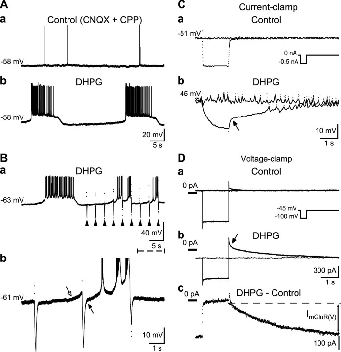
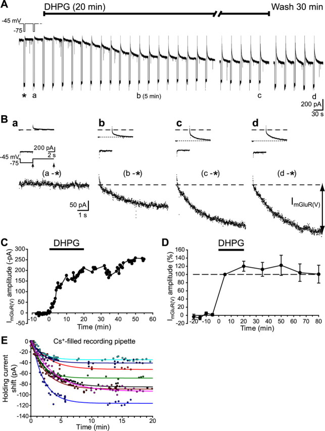
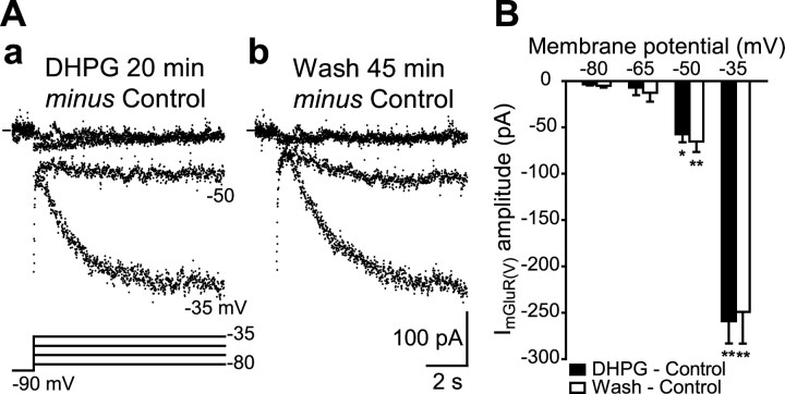
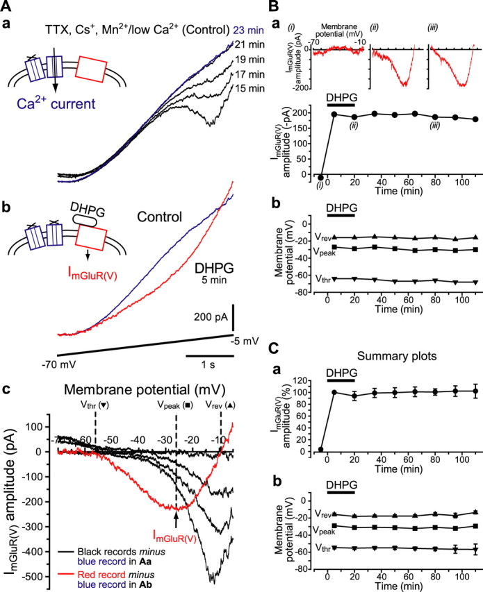
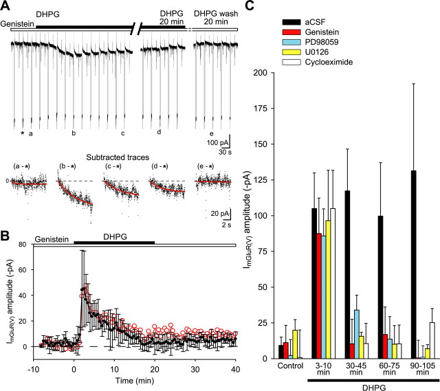
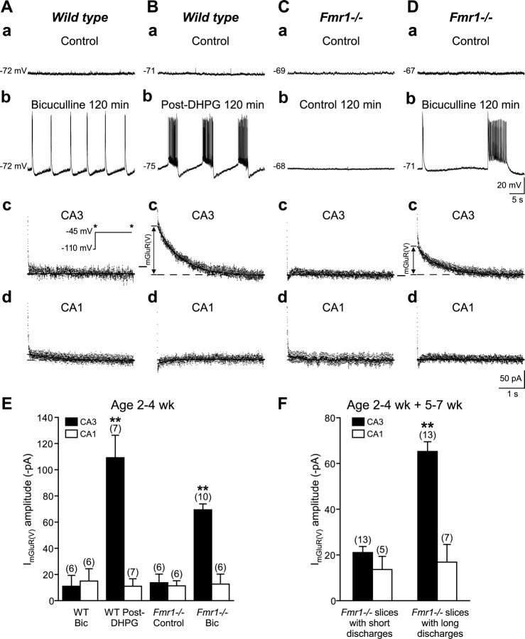
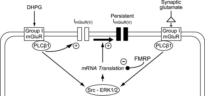
Stimulation of group I metabotropic glutamate receptors (mGluRs) by the agonist (S)-dihydroxyphenylglycine in the hippocampus transforms normal neuronal activity into prolonged epileptiform discharges. The conversion is long lasting in that epileptiform discharges persist after washout of the inducing agonist and serves as a model of epileptogenesis. The group I mGluR model of epileptogenesis took on special significance because epilepsy associated with fragile X syndrome (FXS) may be caused by excessive group I mGluR signaling. At present, the plasticity mechanism underlying the group I mGluR-mediated epileptogenesis is unknown. I(mGluR(V)), a voltage-gated cationic current activated by group I mGluR agonists in CA3 pyramidal cells in the hippocampus, is a possible candidate. I(mGluR(V)) activation is associated with group I mGluR agonist-elicited epileptiform discharges. For I(mGluR(V)) to play a role in epileptogenesis, long-term activation of the current must occur after group I mGluR agonist exposure or synaptic stimulation. We observed that I(mGluR(V)), once induced by group I mGluR agonist stimulation in CA3 pyramidal cells, remained undiminished for hours after agonist washout. In slices prepared from FXS model mice, repeated stimulation of recurrent CA3 pyramidal cell synapses, effective in eliciting mGluR-mediated epileptiform discharges, also induced long-lasting I(mGluR(V)) in CA3 pyramidal cells. Similar to group I mGluR-mediated prolonged epileptiform discharges, persistent I(mGluR(V)) was no longer observed in preparations pretreated with inhibitors of tyrosine kinase, of extracellular signal-regulated kinase 1/2, or of mRNA protein synthesis. The results indicate that I(mGluR(V)) is an intrinsic plasticity mechanism associated with group I mGluR-mediated epileptogenesis.


Figures

Figure 1.
Figure 1.
DHPG induces rhythmic prolonged intrinsic bursts in CA3 pyramidal cells. A, Intracellular records from synaptically isolated CA3 neurons in CNQX and CPP (20 μm each) before (a) and during DHPG (50 μm; b). Membrane potential values are indicated at the beginning of each current-clamp record. B, Application of hyperpolarizing square-wave pulses (−0.6 nA; 100 ms; 0.33 Hz; arrowheads) to the same cell. A segment of the record in a (dashed line) is expanded in b. The spontaneous depolarization preceding the burst (pacemaker potential, hollow arrow) is suppressed by the pulse (filled arrow). Plateau depolarization during spontaneous bursting is maintained by ImGluR(V). During action potential firing, intracellular Ca2+ accumulates (Bianchi et al., 1999), leading to K+ current activation (Bianchi et al., 1999). When cumulative K+ current activation during the burst exceeds the amplitude of ImGluR(V), net membrane current becomes outward, causing hyperpolarization, deactivation of ImGluR(V), and burst termination. C, Voltage responses to a hyperpolarizing pulse (inset) recorded in CNQX and CPP (a) and after addition of DHPG (b). Note that DHPG induced depolarization of the membrane potential above threshold for action potential firing. Action potentials are clipped by digitization. Sustained activation of ImGluR(V) maintained the resting membrane potential at −45 mV. ImGluR(V) was turned off by hyperpolarization and was activated after release of the hyperpolarization (arrow). Thus, non-inactivating ImGluR(V) contributes to the DHPG-induced depolarization of the resting membrane potential. In C and D, responses to pulses are superimposed to baseline records. D, Voltage-clamp records obtained in the same conditions as in C. The net inward current elicited by DHPG after the pulse (Dc; ImGluR(V)) was obtained by subtracting the record in Da from that in Db and was fitted with a single exponential (solid line). The dashed line is the current level extrapolated from the fit at the end of the pulse. Zero current level is indicated for each voltage-clamp record.
Figure 2.
Figure 2.
Long-lasting induction of ImGluR(V) by DHPG. A, Whole-cell patch-clamp recordings of responses of a CA3 pyramidal cell to DHPG. Hyperpolarizing voltage steps (inset) were applied every 30 s throughout the recording. B, Current responses to pulses in A are shown expanded in the insets. The net inward current elicited by DHPG (ImGluR(V)) after the pulses (segment between arrows in the voltage protocol in a) was obtained by subtracting the control response (*) from the corresponding traces. C, Time course of the ImGluR(V) amplitude for the experiment shown in A. D, Summary data of ImGluR(V) amplitude normalized to that recorded at 5 min DHPG in seven cells. E, Shift of holding currents in eight cells recorded with Cs+-filled pipettes and voltage clamped at −50 mV. Time 0 is the onset of successful whole-cell patch recording. Plots were well fitted with single exponentials (R2 > 0.91).
Figure 3.
Figure 3.
Long-lasting ImGluR(V) is activated by depolarization. A, ImGluR(V) amplitude activated by depolarizing voltage steps (bottom) recorded during (a) and 45 min after (b) DHPG washout. Records were obtained by subtraction of indicated current responses. TTX (1 μm) and Cs+ (5 mm) were present throughout the recording, performed with Cs+-filled pipettes. B, Average amplitude of ImGluR(V) activated by depolarizing pulses to the indicated levels during (filled bars) and 45 min after DHPG (hollow bars) in six cells. The amplitude of ImGluR(V) was maintained after DHPG washout (*p < 0.01; **p < 0.05).
Figure 4.
Figure 4.
Activation properties of long-lasting ImGluR(V). A, Voltage-gated calcium currents were suppressed (a) in a solution containing low Ca2+ (0.2 mm), Mn2+ (1 mm), TTX (1 μm), and Cs+ (5 mm). Under this condition (Control), a linear current response was obtained with a depolarizing ramp from −70 mV to −5 mV (Ab, bottom). With the addition of DHPG, an inward “sag” developed in the current response to the same ramp (b). I–V plots (c) were obtained by subtracting the control current response (blue record) from the indicated responses. The I–V plot provided values for the peak amplitude (arrow) and voltage (Vpeak), threshold (Vthr), and reversal potentials (Vrev) of ImGluR(V). B, Plots of ImGluR(V) amplitude (a), and of Vpeak, Vthr, and Vrev (b), for the current (inset) recorded before (i), during (ii), and after (iii) DHPG application in a CA3 pyramidal cell. C, Summary data from 11 CA3 pyramidal cells.
Figure 5.
Figure 5.
Long-lasting ImGluR(V) is not blocked by the competitive mGluR antagonist MCPG. A, Currents were elicited by depolarizing steps from −80 to −45 mV. The currents shown were obtained by subtracting the corresponding control (i.e., before DHPG application) traces from the responses recorded in the indicated conditions (a, DHPG: after 5 min application of DHPG 50 μm; b, DHPG plus MCPG: after 5 min DHPG 50 μm applied in the presence of MCPG 250 μm; c, DHPG wash: after 30 min of DHPG washout; and c, DHPG wash plus MCPG: after application of MCPG 250 μm for 40 min subsequent to DHPG washout, in the same group of cells shown as DHPG wash). As in Figure 1Dc, subtracted traces were fitted with single exponentials (solid lines). B, Summary data of ImGluR(V) amplitude in the different series of experiments indicated. Significant activation of ImGluR(V) (DHPG, −125.2 ± 21.6 pA vs Control, −5.2 ± 1.1 pA; n = 6; **p < 0.01, Student's paired t test) was prevented in the presence of MCPG 250 μm (DHPG plus MCPG, −4.6 ± 2.7 pA vs Control, −4.1 ± 1.0 pA; n = 5; p = 0.90). In contrast, ImGluR(V) persisting after DHPG washout was not affected by the same concentration of the antagonist (DHPG wash plus MCPG, −107.8 ± 31.5 pA vs DHPG wash, −108.8 ± 28.9 pA; n = 6; p = 0.85).
Figure 6.
Figure 6.
Long-lasting ImGluR(V) is suppressed by inhibitors of tyrosine kinase, ERK1/2, and protein synthesis. A, Current responses of a CA3 pyramidal cell to hyperpolarizing voltage steps from −75 to −45 mV (downward deflections) before, during (filled bar), and after DHPG in the continuous presence of the tyrosine kinase inhibitor genistein (30 μm; hollow bar). ImGluR(V) elicited after the hyperpolarizing pulse was obtained by subtracting the control response (*) from the corresponding indicated responses (Subtracted traces). Red lines are single exponential fits. B, Time course of ImGluR(V) amplitude for the cell shown in A (red circles) and summary data plot for five cells (filled circles). C, Mean ImGluR(V) amplitude recorded in control and in DHPG (aCSF; n = 8), in DHPG plus genistein (n = 4), in DHPG plus PD98059 (n = 3), in DHPG plus U0126 (n = 3), in DHPG plus cycloheximide (n = 4). In each experiment, agents were added 60 min before DHPG. Peak ImGluR(V) activation was observed in all experimental conditions with blockers after 3–10 min of DHPG application.
Figure 7.
Figure 7.
Agonist-induced or synaptically induced ImGluR(V) underlies prolonged epileptiform discharges. A, Intracellular recordings from a CA3 pyramidal cell in control solution (aCSF; a) and 120 min after addition of bicuculline 50 μm (b). The burst firing represents short duration (300–500 ms) synchronized discharges of the CA3 population. The same cell was recorded in CNQX and CPP after the bicuculline treatment (c). ImGluR(V) amplitude at −45 mV, activated after a hyperpolarizing pulse to −110 mV, was measured under voltage clamp. In this condition, ImGluR(V) was negligible. A CA1 cell in the same slice was recorded under the same condition (d). B, Prolonged epileptiform discharges induced by DHPG persisted after agonist washout (b). Addition of CNQX and CPP blocked the synchronized discharges. ImGluR(V) amplitude activated in the same cell was measured under voltage clamp (c). A CA1 cell in the same slice was recorded under the same condition (d). C and D, CA3 pyramidal cells from Fmr1−/− mice. C, In aCSF, ImGluR(V) was negligible in both CA3 (c) and CA1 (d) cells. D, Bicuculline induced group I mGluR-mediated prolonged epileptiform discharges (b). In the same cell, recorded in CNQX and CPP to suppress the epileptiform discharges, ImGluR(V) was observed in CA3 (c) but not in CA1 (d) cells. E, Summary data of ImGluR(V) amplitude under the different conditions in (A–Dc,d). Data are from 2- to 4- week-old (young) mice. Numbers of cells are indicated in parenthesis (**p < 0.01). F, Mean ImGluR(V) amplitude in CA3 and CA1 cells recorded in slices isolated from Fmr1−/− mice that showed interictal (short; <1.5 s) and prolonged epileptiform (long; ≥1.5 s) discharges. Data are pooled from young and older (5–7 week) mice. Bic, Bicuculline.
Figure 8.
Figure 8.
Schematic representation of the effects of group I mGluR stimulation in hippocampal CA3 pyramidal cells. Group I mGluRs activated by DHPG and by synaptic glutamate are depicted separately for clarity. DHPG is likely to activate extrasynaptic and synaptic group I mGluRs. Model of persistent activation of ImGluR(V): DHPG action (left side of the scheme). DHPG activates ImGluR(V) via stimulation of mGluR1 and mGluR5 through PLCβ1-mediated signaling (Chuang et al., 2001). Stimulation of group I mGluRs also activates protein synthesis via Src tyrosine kinase-ERK1/2 signaling-dependent mRNA translation. The group I mGluR-mediated protein synthesis is required to induce long-lasting ImGluR(V) (thick arrow). FMRP regulation of synaptic group I mGluR-mediated mRNA translation: Synaptic glutamate (right side of the scheme). Synaptic stimulation of group I mGluRs is elicited by spontaneous synchronized discharges of CA3 pyramidal cells in the presence of bicuculline (Chuang et al., 2005). Prolonged periods of synaptic stimulation of group I mGluRs induced persistent ImGluR(V) in Fmr1−/− preparations, whereas similar pattern of stimulation was ineffective in ImGluR(V) activation in wild-type preparations. Presumably, ImGluR(V) induction is prevented by FMRP in the wild-type preparation. Whereas synaptic induction of ImGluR(V) is prevented in the wild type by FMRP, we note that DHPG is effective in the induction of ImGluR(V) in the same preparation. There are at least two possible explanations for the difference in synaptic- versus agonist-induced responses in the wild-type preparation. (1) Only synaptic group I mGluRs are regulated by FMRP, whereas DHPG activates also nonsynaptic group I mGluRs which are not subject to FMRP regulation. Evidence for the inhibitory role of FMRP on synaptically activated prolonged epileptiform discharges is from data in Figure 7, showing that synaptic activation of group I mGluRs can elicit epileptogenesis in the absence of FMRP (Fmr1−/− preparations) but not in the presence of functional FMRP (wild-type preparations). (2) Stimulation of group I mGluRs by DHPG leads to downregulation of FMRP function, possibly via activation of the ubiquitin-proteasome system (Hou et al., 2006), whereas synaptic stimulation does not affect FMRP levels.

References

    1. Aschrafi A, Cunningham BA, Edelman GM, Vanderklish PW. The fragile X mental retardation protein and group I metabotropic glutamate receptors regulate levels of mRNA granules in brain. Proc Natl Acad Sci U S A. 2005;102:2180–2185. - PMC - PubMed
    1. Bear MF, Huber KM, Warren ST. The mGluR theory of fragile X mental retardation. Trends Neurosci. 2004;27:370–377. - PubMed
    1. Bianchi R, Wong RKS. Excitatory synaptic potentials dependent on metabotropic glutamate receptor activation in guinea-pig hippocampal pyramidal cells. J Physiol. 1995;487:663–676. - PMC - PubMed
    1. Bianchi R, Young SR, Wong RKS. Group I mGluR activation causes voltage-dependent and -independent Ca2+ rises in hippocampal pyramidal cells. J Neurophysiol. 1999;81:2903–2913. - PubMed
    1. Bianchi R, Chuang SC, Wong RKS. Pharmacology of a slowly inactivating outward current in hippocampal CA3 pyramidal neurons. J Neurophysiol. 2006;96:1116–1123. - PubMed

Publication types

MeSH terms

Substances