

Peripheral T-cell lymphomas with a follicular growth pattern are derived from follicular helper T cells (TFH) and may show overlapping features with angioimmunoblastic T-cell lymphomas
- PMID: 19295409
- PMCID: PMC4838638
- DOI: 10.1097/PAS.0b013e3181971591
Peripheral T-cell lymphomas with a follicular growth pattern are derived from follicular helper T cells (TFH) and may show overlapping features with angioimmunoblastic T-cell lymphomas
Abstract
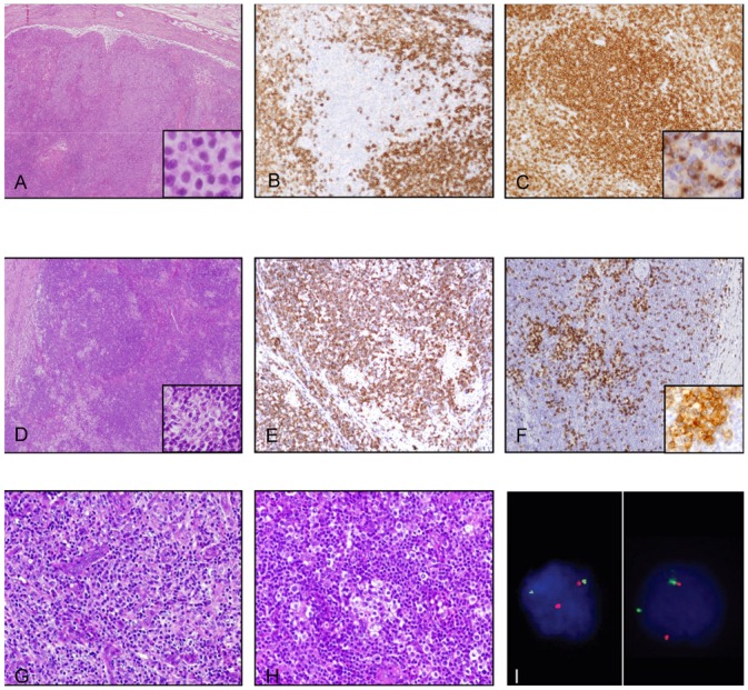
Rare cases of peripheral T-cell lymphomas with follicular growth pattern (PTCL-F) have been recently reported, and their association with t(5;9)(q33;q22) involving ITK and SYK has been suggested. However, the clinicopathologic aspects of PTCL-F are poorly described and the normal cell counterpart of this subgroup of lymphoma is still unknown. Therefore, we analyzed the pathologic, phenotypic, and cytogenetic features of a series of 30 patients (range: 33 to 88 y) that showed histopathologic features of PTCL-F in at least 1 biopsy (n=30), either at initial presentation (n=26) or at relapse (n=4). Neoplastic cells were medium-sized clear cells that were CD4+ (24/27, 89%), CD10+ (21/29, 72%), BCL-6+ (14/19, 74%), and expressed programed death-1 (27/27, 100%), CXCL13 (23/27, 85%), and ICOS (11/11, 100%), markers of follicular helper T cells (TFH). Four of 22 patients (18%) had t(5;9)(q33;q22) detected by fluorescence in situ hybridization. Patients with clinical data available had multiple lymphadenopathies (25/28, 89%), stage III to IV diseases (17/26, 65%), B symptoms (7/27, 26%), and skin lesions (6/23, 26%). Three patients with sequential biopsies disclosed clinical and histopathologic features of angioimmunoblastic T-cell lymphoma at initial presentation. Our results show that this rare form of PTCL-F (1) has an immunophenotype indicative of derivation from TFH cells, (2) is associated with t(5;9) in a proportion of cases, and (3) shows some overlapping features with angioimmunoblastic T-cell lymphoma, raising the question of a possible relationship.
Figures
References
-
- Attygalle A, Al-Jehani R, Diss TC, et al. Neoplastic T cells in angioimmunoblastic T-cell lymphoma express CD10. Blood. 2002;99:627–633. - PubMed
-
- Attygalle AD, Chuang SS, Diss TC, et al. Distinguishing angioimmunoblastic T-cell lymphoma from peripheral T-cell lymphoma, unspecified, using morphology, immunophenotype and molecular genetics. Histopathology. 2007;50:498–508. - PubMed
-
- Bacon CM, Paterson JC, Liu H, et al. Peripheral T-cell lymphoma with a follicular growth pattern: derivation from follicular helper T cells and relationship to angioimmunoblastic T-cell lymphoma. Br J Haematol. 2008 doi: 10.1111/j.1365-2141.2008.07352.x. published online on August 22 2008. - DOI - PubMed
-
- Bolen JB, Brugge JS. Leukocyte protein tyrosine kinases: potential targets for drug discovery. Annu Rev Immunol. 1997;15:371–404. - PubMed
Publication types
MeSH terms
Substances
LinkOut - more resources
Full Text Sources
Research Materials
Miscellaneous
