. 2009 Mar 19;458(7236):299-304.
doi: 10.1038/nature07842.

Activation of CaMKII in single dendritic spines during long-term potentiation

Affiliations

Activation of CaMKII in single dendritic spines during long-term potentiation

Seok-Jin R Lee et al. Nature. .

Abstract

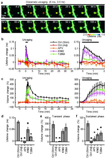
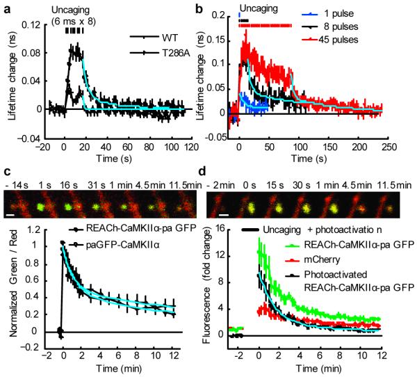
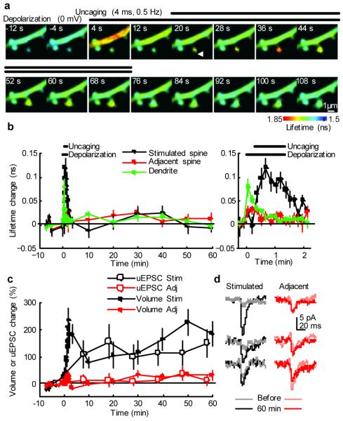
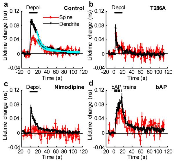
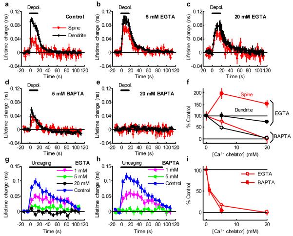
Calcium/calmodulin-dependent kinase II (CaMKII) plays a central part in long-term potentiation (LTP), which underlies some forms of learning and memory. Here we monitored the spatiotemporal dynamics of CaMKII activation in individual dendritic spines during LTP using two-photon fluorescence lifetime imaging microscopy, in combination with two-photon glutamate uncaging. Induction of LTP and associated spine enlargement in single spines triggered transient ( approximately 1 min) CaMKII activation restricted to the stimulated spines. CaMKII in spines was specifically activated by NMDA receptors and L-type voltage-sensitive calcium channels, presumably by nanodomain Ca(2+) near the channels, in response to glutamate uncaging and depolarization, respectively. The high degree of compartmentalization and channel specificity of CaMKII signalling allow stimuli-specific spatiotemporal patterns of CaMKII signalling and may be important for synapse-specificity of synaptic plasticity.


Figures

Figure 1
Figure 1. Simultaneous measurements of CaMKII activation and structural plasticity in single spines using 2pFLIM combined with 2-photon glutamate uncaging
a, Fluorescence lifetime images of Green-Camuiα during the induction of spine structural plasticity by 2-photon glutamate uncaging in the absence of extracellular Mg2+. Longer lifetimes imply increased activity. The white arrowhead indicate the location of uncaging laser spot. b, Averaged time course of fluorescence lifetime change of Green-Camuiα in the stimulated spine (indicated as “Stim”) and adjacent spines (“Adj”, within 5 μm of stimulated spine). Data using pharmacological inhibitors and T286A mutant Green-Camuiα are also shown. The number of samples (spines / neurons) is 35 / 30 for stimulated spines, 29 / 28 for adjacent spines, 5 / 4 for AP5, 7 / 5 for KN62 and 18 / 11 T286A. Right panel: closer view of fluorescence lifetime change during uncaging. c, Averaged time course of spine volume change in stimulated spines and adjacent spines. Right: closer view of the volume change. d, CaMKII activation (averaged over 0 - 40 s or first 5 points). Stars denote statistical significance between control and others (p < 0.05). e, Transient volume change (volume change averaged over 1.5 - 2 min subtracted by that averaged over 25 - 30 min) . f, Sustained volume change (volume change averaged over 25 - 30 min).
Figure 2
Figure 2. Measurements of parameters that determine spine-specificity: inactivation kinetics and mobility of CaMKII
a, Measurements of CaMKII activation and inactivation following brief stimulation (8 pulses, 0.5 Hz) of spines using wild type and T286A Green Camuiα in zero extracellular Mg2+. These experiments were performed in an interleaved manner. The solid cyan curve indicates double exponential functions obtained by fitting to wild type data (time constants: 6 s [80%] and 45 s [20%]). The dashed cyan curve indicates single exponential function with 1 s time constant. The number of samples (spines / neurons) is 25 / 5 for wild type and 28 / 4 for T286A. b. CaMKII inactivation kinetics following different number of uncaging pulses (1, 8 and 45 pulses, 0.5 Hz). Data were fitted with double exponential functions (cyan curves). The time constants were 9.3 s (71%) and > 100 s (29 %) for 1 pulse, 4.7 s (61 %) and 136.6 s (39 %) for 8 pulses, 4.5 s (61 %) and 102 s (39 %) for 45 pulses. The number of samples (spines / neurons) is 28 / 9 (1 pulses), 16 / 8 (8 pulses) and 13 / 7 (45 pulses). c, Measurements of spine-dendrite coupling of CaMKII using photoactivatable GFP (paGFP). We used Green-Camuiα with mEGFP replaced by paGFP (REACh-CaMKIIα-paGFP) or paGFP-CaMKIIα. Top panel: Green and red fluorescence before and after photoactivation in a neuron expressing REACh-CaMKIIα-paGFP (green) and mCherry (red). Bar, 1 μm. Bottom panel: Averaged time course of the ratio of red and green. Signal was normalized to the peak fluorescence change. The first point is 1 s after the photoactivation. By fitting with double exponential functions, the decay time constants was obtained as 1.3 min (48 %) and 17.0 min (52 %) for REACh-CaMKIIα-paGFP (cyan solid curve), and 1.0 minutes (53 %) and 20.7 minutes (47 %) for paGFP-CaMKIIα (cyan dashed curve). The number of samples (spines / neurons) is 15 / 5 (REACh-CaMKIIα-paGFP) and 22 / 9 (paGFP-CaMKIIα). d. Spine-dendrite coupling of Green-Camuiα during structural plasticity. During glutamate uncaging (720 nm, 6 ms, 45 pulses in Mg2+ free solution), REACh-CaMKIIα-paGFP can be photoactivated. We subtracted the normalized red fluorescence from the normalized green fluorescence, assuming that the small baseline fluorescence of REACh-CaMKIIα-paGFP behaves similarly to mCherry fluorescence change (see Supplementary Fig. 3). By fitting with double exponential functions (cyan curve), the decay time constants were obtained as 1.6 min (82 %) and 15.3 min (18 %). The number of samples (spines / neurons) is 10 / 7.
Figure 3
Figure 3. Imaging CaMKII activation during LTP induction by 2-photon glutamate uncaging paired with postsynaptic depolarization
a, Fluorescence lifetime images of Green-Camuiα during the induction of LTP by 2-photon glutamate uncaging paired with postsynaptic depolarization. b, Averaged time course of fluorescence lifetime change of Green-Camuiα in the stimulated spine, adjacent spines (within 5 μm of stimulated spine), and dendritic shaft close to the stimulated spine. Right: closer view of fluorescence lifetime change during uncaging. Average of 8 stimulated spines, 13 adjacent spines and 8 dendrites from 8 neurons. Plot of stimulated spines is noisier due to smaller number of samples. Plots of stimulated spines in b - d are from the same spines. c, Averaged time course of changes in spine volume and the amplitude of uncaging-evoked EPSC at -65 mV (uEPSC) in the stimulated spine and adjacent spines (within 5 μm). For uEPSC, each point is average of four trials at 5 s intervals. Uncaging spot was relocated to the tip of spines before each four trial measurement. d, uEPSC at single spines before and 60 minutes after LTP induction (average of 4 trials, filtered with 2 ms window).
Figure 4
Figure 4. Differential activation of CaMKII in spines and dendrites by postsynaptic depolarization
a, High temporal resolution measurements of the fluorescence lifetime change of Green-Camuiα in spines and dendrites in response to postsynaptic depolarization (0 mV, 16 s, indicated by the bar) in spines (red) and dendritic shafts (black). Average of 53 spines and 30 dendrites from 12 neurons. The cyan lines are a double exponential function obtained by fitting. The time constants are 11.3 s (84 %) and > 300 s (16%) for spines, and 6.8 s (60 %) and 43.7 s (40 %) for dendrites. b, Fluorescence lifetime change of T286A mutant of Green-Camuiα. Average of 19 spines and 14 dendrites from 8 neurons. c, Fluorescence lifetime change of Green-Camuiα in the presence of L-type VSCC inhibitor (20 μM Nimodipine). Average of 22 spines and 15 dendrites from 7 neurons. d, Bursts of back-propagating action potentials (bAP) induced CaMKII activation at proximal dendrites (< 100 μm). We applied 8 bAP bursts, each 83 Hz for 0.5 s, at 2 s intervals. Average of 22 spines and 12 dendrites from 7 neurons).
Figure 5
Figure 5. Effects of Ca2+ chelators on CaMKII activation
a-e, Fluorescence lifetime change of Green-Camuiα in spines and dendrites in response to postsynaptic depolarization (0 mV, 0 - 16 s, indicated by the black bar) in spines (red) and dendritic shafts (black) in neurons patch-clamped with electrodes containing no Ca2+ chelator (a), 5 mM EGTA (b), 20 mM EGTA (c), 5 mM BAPTA (d) or 20 mM BAPTA (e). Number of samples (spines / dendrites / neurons) is 18 / 11 / 6 for control (a), 29 / 14 / 7 for 5 mM EGTA (b), 15 / 7 / 4 for 20 mM EGTA (c), 21 / 7 / 5 for 5 mM BAPTA (d) and 18 / 7 / 4 for 20 mM BAPTA (e). Imaging was performed more than 15 min after establishing whole-cell patch. f, Dependence of fluorescence lifetime change (averaged over 0 - 16 s) by depolarization on Ca2+ chelator concentration (EGTA: closed circle, BAPTA: open circle, red: spine, black: dendrite). Fluorescence lifetime was normalized to control. g-h, Fluorescence lifetime change of Green-Camuiα in response to glutamate uncaging (6 ms, 45 stimuli in zero extracellular Mg2+) in neurons patch-clamped with electrodes containing 1 - 20 mM EGTA (g), 1 - 5 mM BAPTA (h) in a current-clamp mode. Controls are measured in the same neurons before patch-clamp. Patch-clamping with internal solution without Ca2+ chelator did not alter fluorescence lifetime change (Supplementary Fig. 7). Number of samples (spines / neurons) is 22 / 9 for control in g, 12 / 3 for 1 mM EGTA, 15 / 3 for 5 mM EGTA, 17 / 3 for 20 mM EGTA, 16 / 7 for control in h, 17 / 4 for 1 mM BAPTA, 14 / 3 for 5 mM BAPTA. i, Dependence of fluorescence lifetime change (averaged over 0 - 40 s) induced by glutamate uncaging on Ca2+ chelator concentration (EGTA: open circle, BAPTA: closed circle). Fluorescence lifetime was normalized to control.

Comment in

References

    1. Rosenberg OS, et al. Oligomerization states of the association domain and the holoenyzme of Ca2+/CaM kinase II. Febs J. 2006;273:682–694. - PubMed
    1. Lisman JE, Zhabotinsky AM. A model of synaptic memory: a CaMKII/PP1 switch that potentiates transmission by organizing an AMPA receptor anchoring assembly. Neuron. 2001;31:191–201. - PubMed
    1. Merrill MA, Chen Y, Strack S, Hell JW. Activity-driven postsynaptic translocation of CaMKII. Trends Pharmacol Sci. 2005;26:645–653. - PubMed
    1. Hudmon A, et al. CaMKII tethers to L-type Ca2+ channels, establishing a local and dedicated integrator of Ca2+ signals for facilitation. J Cell Biol. 2005;171:537–547. - PMC - PubMed
    1. Takao K, et al. Visualization of synaptic Ca2+ /calmodulin-dependent protein kinase II activity in living neurons. J Neurosci. 2005;25:3107–3112. - PMC - PubMed

Publication types

MeSH terms