. 2009 Sep;129(9):2175-83.
doi: 10.1038/jid.2009.65. Epub 2009 Mar 19.

Th17 cytokines stimulate CCL20 expression in keratinocytes in vitro and in vivo: implications for psoriasis pathogenesis

Affiliations

Th17 cytokines stimulate CCL20 expression in keratinocytes in vitro and in vivo: implications for psoriasis pathogenesis

Erin G Harper et al. J Invest Dermatol. 2009 Sep.

Abstract

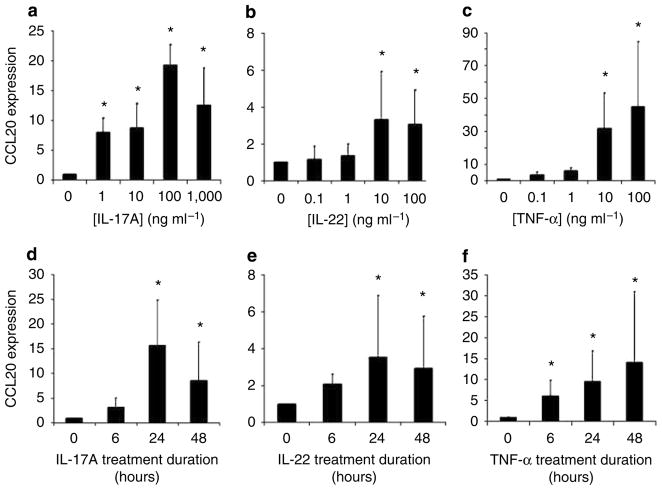
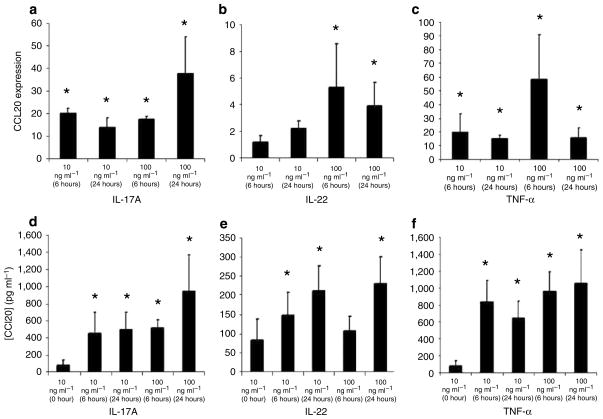
T helper (Th) 17 cells have recently been implicated in psoriasis pathogenesis, but mechanisms of how these cells traffic into inflamed skin are unknown. By immunostaining for interleukin (IL)-17A and IL-22, we show numerous cells present in psoriasis lesions that produce these cytokines. We next found that Th17 cytokines (IL-17A, IL-22, and tumor necrosis factor (TNF)-alpha) markedly increased the expression of CC chemokine ligand (CCL) 20, a CC chemokine receptor (CCR)6 ligand, in human keratinocyte monolayer and raft cultures in a dose- and time-dependent manner. Lastly, we showed in mice that subcutaneous injection with recombinant IL-17A, IL-22, or TNF-alpha led to the upregulation of both CCL20 and CCR6 expression in skin as well as cutaneous T-cell infiltration. Taken together, these data show that Th17 cytokines stimulate CCL20 production in vitro and in vivo, and thus provide a potential explanation of how CCR6-positive Th17 cells maintain their continual presence in psoriasis through a positive chemotactic feedback loop.


Conflict of interest statement

CONFLICT OF INTEREST

The authors state no conflict of interest.

Figures

Figure 1
Figure 1. IL-17A-positive and IL-22-positive cells are abundant in psoriasis lesional skin
Formalin-fixed paraffin-embedded tissue sections obtained from seven psoriasis patients and three healthy individuals were examined by IHC. Tissue was stained with either anti-CD3 or anti-IL-17A antibodies. In six separate individuals with psoriasis, lesional skin was biopsied and snap frozen. Fresh-frozen sections were stained with either anti-CD3 or anti-IL-22 antibodies. Increased numbers of IL-17A- and IL-22-expressing cells (brown or purple in color) were present in lesional skin when compared with healthy skin. Representative photos are shown. Bar = 10 μm in figures labeled × 40, 20 μm in figures labeled × 20, and 40 μm in figures labeled × 10.
Figure 2
Figure 2. IL-17A, IL-22, and TNF-α increase CCL20 mRNA expression by normal human KC in a dose- and time-dependent manner in vitro
Cells were treated with the indicated concentrations of (a) IL-17A, (b) IL-22, or (c) TNF-α for 24 hours. Cells were treated with the optimal cytokine concentrations of (d) IL-17A, (e) IL-22, or (f) TNF-α for 6, 24, or 48 hours. mRNA was harvested and CCL20 transcripts were quantified by real-time RT-PCR analyses for all experiments. All experiments were performed in triplicate at least three separate times and the average mRNA increase and SD is reported. Significant differences were detected using the Mann–Whitney unpaired two-tailed t-test (*Indicates significance, P<0.05).
Figure 3
Figure 3. IL-17A, IL-22, and TNF-α increase CCL20 protein expression by normal human KC in a dose- and time-dependent manner in vitro
Cells were treated with the indicated concentrations of (a) IL-17A, (b) IL-22, or (c) TNF-α for 24 hours. Cells were treated with the optimal cytokine concentrations of (d) IL-17A, (e) IL-22, or (f) TNF-α for 6, 24, or 48 hours. Cell-free supernatants were harvested and CCL20 protein levels were quantified by ELISA for all experiments. All experiments were performed in triplicate at least three separate times and the average protein levels and SD are reported. Significant differences were detected using the Mann–Whitney unpaired two-tailed t-test (*Indicates significance, P<0.05).
Figure 4
Figure 4. IL-17A, IL-22, and TNF-α increase CCL20 mRNA and protein expression by RHE in a dose- and time-dependent manner
Stratified KC in RHE were treated with the indicated concentrations of (a) IL-17A, (b) IL-22, or (c) TNF-α for 6 or 24 hours. Total RNA was harvested and CCL20 transcripts were quantified by real-time RT-PCR analyses. RHE were treated with the optimal cytokine concentrations of (d) IL-17A, (e) IL-22, or (f) TNF-α for 6 or 24 hours. Cell-free supernatants were harvested and CCL20 protein levels were quantified by ELISA. All experiments were performed in triplicate at least three separate times and the average protein levels and SD are reported. Significant differences were detected using the Mann–Whitney unpaired two-tailed t-test (*Indicates significance, P<0.05).
Figure 5
Figure 5. CCL20 and CCR6 upregulation and T-cell infiltration in murine skin injected with Th17 cytokines
(ac) Balb/c mouse ears were injected with 500 ng of IL-17A, IL-22, TNF-α, or PBS daily for 5 days. (a and c) CCL20 and CCR6 mRNA expression levels in injected ears were determined by real-time RT-PCR. Significant differences were detected using the Mann–Whitney unpaired two-tailed t-test (*Indicates significance, P<0.05). (b) CCL20 protein expression was measured by ELISA. Significant differences were detected using the unpaired t-test with Welch’s correction (*Indicates significance, P<0.05). (ac) Data from 10 mice in each treatment group are shown. (d) Representative histologic photos showing CD3 positive T cells in murine skin injected with Th17 cytokines. Bar= 40 μm.

References

    1. Acosta-Rodriguez EV, Napolitani G, Lanzavecchia A, Sallusto F. Interleukins 1beta and 6 but not transforming growth factor-beta are essential for the differentiation of interleukin 17-producing human T helper cells. Nat Immunol. 2007a;8:942–9. - PubMed
    1. Acosta-Rodriguez EV, Rivino L, Geginat J, Jarrossay D, Gattorno M, Lanzavecchia A, et al. Surface phenotype and antigenic specificity of human interleukin 17-producing T helper memory cells. Nat Immunol. 2007b;8:639–46. - PubMed
    1. Annunziato F, Cosmi L, Santarlasci V, Maggi L, Liotta F, Mazzinghi B, et al. Phenotypic and functional features of human Th17 cells. J Exp Med. 2007;204:1849–61. - PMC - PubMed
    1. Austin LM, Ozawa M, Kikuchi T, Walters IB, Krueger JG. The majority of epidermal T cells in Psoriasis vulgaris lesions can produce type 1 cytokines, interferon-gamma, interleukin-2, and tumor necrosis factor-alpha, defining TC1 (cytotoxic T lymphocyte) and TH1 effector populations: a type 1 differentiation bias is also measured in circulating blood T cells in psoriatic patients. J Invest Dermatol. 1999;113:752–9. - PubMed
    1. Blauvelt A. New concepts in the pathogenesis and treatment of psoriasis: key roles for IL-23, IL-17A and TGF-b1. Expert Rev Dermatol. 2007;2:1–10.

Publication types