

Is scleroderma a vasculopathy?
- PMID: 19296882
- PMCID: PMC4091992
- DOI: 10.1007/s11926-009-0015-3
Is scleroderma a vasculopathy?
Abstract
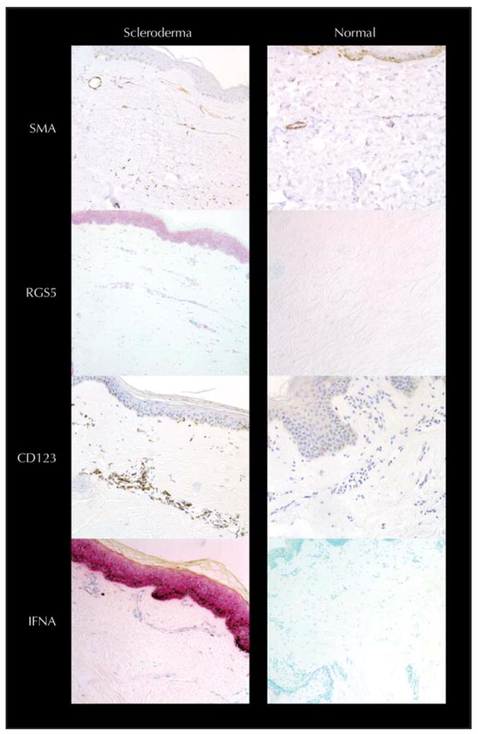
Described as an autoimmune collagen vascular disease, the most striking feature of scleroderma may be a systemic vasculopathy. This vasculopathy includes characteristic noninflammatory macrovascular and microvascular changes with dramatic and possibly occlusive formation of a thickened neointima. Scleroderma vessels also have an unusual endothelial phenotype, with loss of normal markers including vascular endothelial (VE)-cadherin. These endothelial cells express type 1 interferon and regulator of G protein signaling 5 (RGS5), two molecules associated with vascular rarefaction. These genes may be important because tissue is hypoxic with high levels of vascular endothelial growth factor (VEGF), especially early in the disease. The combination of VEGF and rarefaction is not necessarily paradoxical. VEGF-mediated angiogenesis creates labile vessels that may not survive unless the vessel acquires a smooth muscle coat. The combination of interferon and RGS5 is consistent with an antiangiogenic phenotype. We offer a hypothesis that places vascular injury at the center of this disease and also suggest possible clinical approaches for arresting and/or reversing the disease.
Conflict of interest statement
Disclosures
No potential conflicts of interest relevant to this article were reported.
Figures
References
-
- LeRoy EC. Systemic sclerosis. A vascular perspective. Rheum Dis Clin North Am. 1996;22:675–694. - PubMed
-
- Maricq HR. Capillary abnormalities, Raynaud’s phenomenon, and systemic sclerosis in patients with localized scleroderma. Arch Dermatol. 1992;128:630–632. - PubMed
-
- Carpentier PH, Satger B, Poensin D, Maricq HR. Incidence and natural history of Raynaud phenomenon: a long-term follow-up (14 years) of a random sample from the general population. J Vasc Surg. 2006;44:1023–1028. This article demonstrates that, although Raynaud’s phenomenon does tend to be a prodrome for scleroderma, it is not practical to follow these patients over time. - PubMed
-
- Iwata H, Sata M. Potential contribution of bone marrow-derived precursors to vascular repair and lesion formation: lessons from animal models of vascular diseases. Front Biosci. 2007;12:4157–4167. This article follows the lineage and contribution of cells in the blood vessel wall. - PubMed
-
- Passman JN, Dong XR, Wu SP. A Sonic hedgehog signaling domain in the arterial adventitia supports resident sca1+ smooth muscle progenitor cells. Proc Natl Acad Sci U S A. 2008;105:9349–9354. This article and the articles by Carpentier et al. [3•] and Iwata and Sata [4•] provide recent evidence that Sonic hedgehog maintains vasculature in static state and that perturbation of signaling is associated with vascular remodeling. - PMC - PubMed
Publication types
MeSH terms
Substances
Grants and funding
LinkOut - more resources
Full Text Sources
Medical