. 2009 Jul;21(7):1169-79.
doi: 10.1016/j.cellsig.2009.03.006. Epub 2009 Mar 16.

Arf GAP2 is positively regulated by coatomer and cargo

Affiliations

Arf GAP2 is positively regulated by coatomer and cargo

Ruibai Luo et al. Cell Signal. 2009 Jul.

Abstract

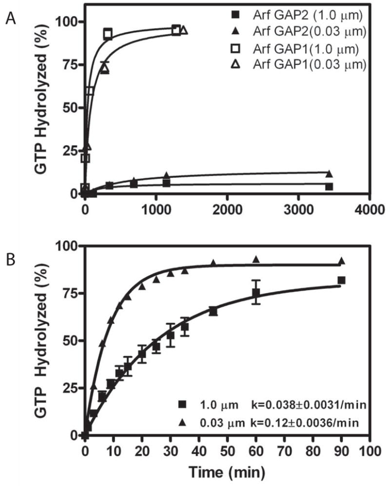
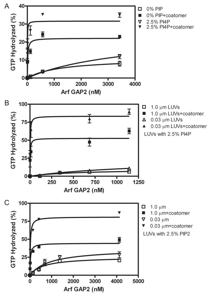
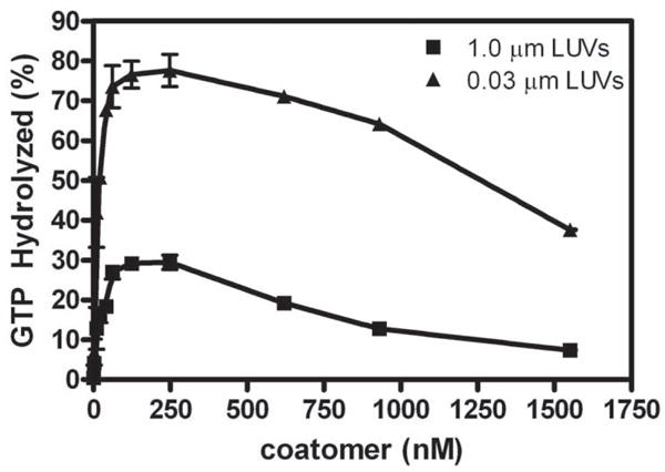
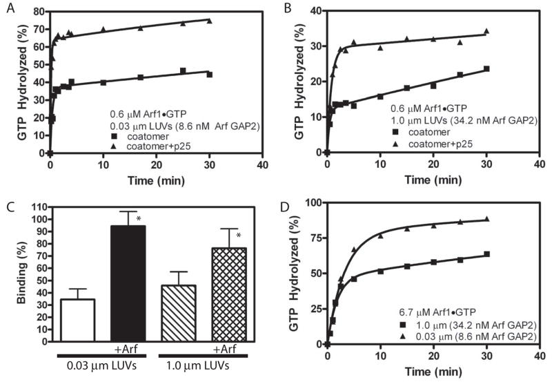
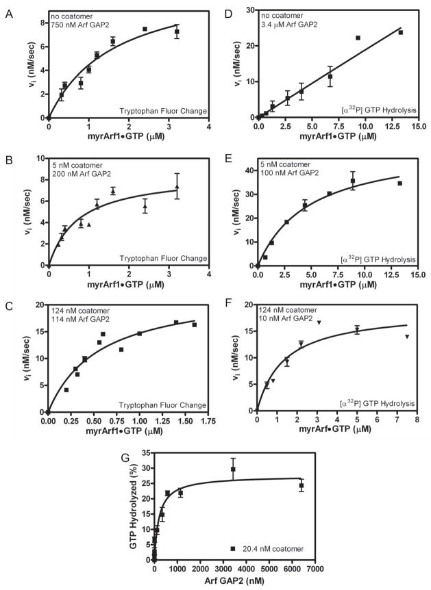
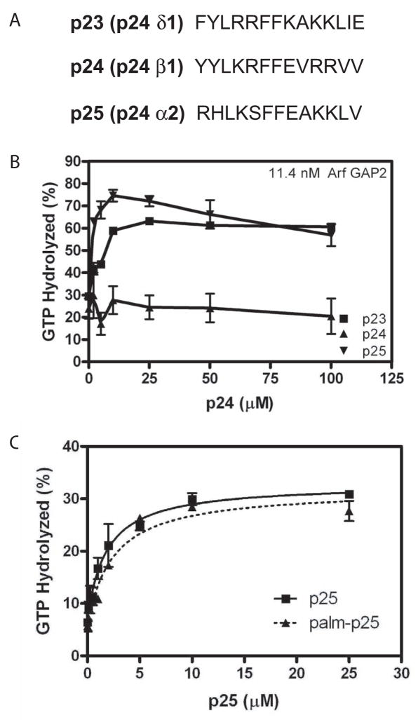
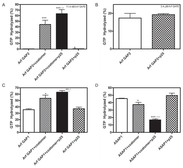
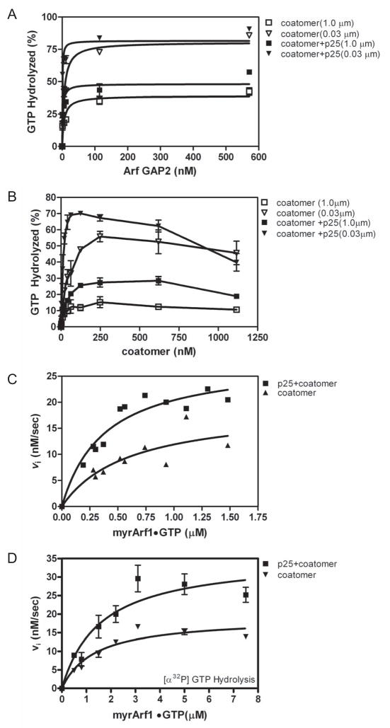
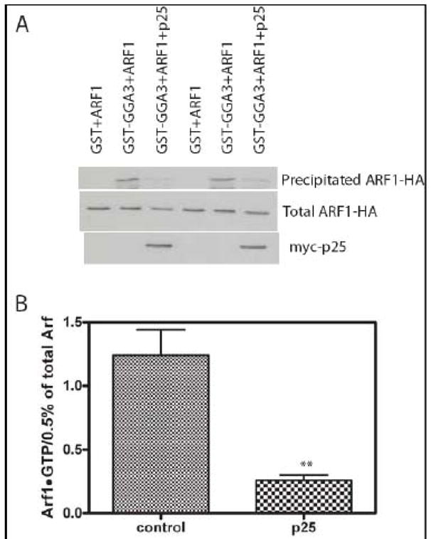
Arf GAP2 is one of four Arf GAPs that function in the Golgi apparatus. We characterized the kinetics of Arf GAP2 and its regulation. Purified Arf GAP2 had little activity compared to purified Arf GAP1. Of the potential regulators we examined, coatomer had the greatest effect, stimulating activity one to two orders of magnitude. The effect was biphasic, with half-maximal activation observed at 50 nM coatomer and activation peaking at approximately 150 nM coatomer. Activation by coatomer was greater for Arf GAP2 than has been reported for Arf GAP1. The effects of phosphoinositides and changes in vesicle curvature on GAP activity were small compared to coatomer; however, both increased coatomer-dependent activity. Peptides from p24 cargo proteins increased Arf GAP2 activity by an additional 2- to 4-fold. The effect of cargo peptide was dependent on coatomer. Overexpressing the cargo protein p25 decreased cellular Arf1*GTP levels. The differential sensitivity of Arf GAP1 and Arf GAP2 to coatomer could coordinate their activities. Based on the common regulatory features of Arf GAP1 and 2, we propose a mechanism for cargo selection in which GTP hydrolysis triggered by cargo binding to the coat protein is coupled to coat polymerization.


Figures

Figure 1
Figure 1. Arf GAP2 activity
GAP activity using myrArf1•GTP as a substrate was determined in the presence of LUVs formed by extrusion through membranes with pores of the indicated diameters. Total phospholipid concentration was 500 μM. A. Comparison of recombinant purified Arf GAP1 and Arf GAP2. [1–521] Arf GAP2 His6 and [1–415]Arf GAP1-His6 were titrated into a reaction containing [α32P]GTP•myrArf1. The fraction of GTP converted to GDP in 3 min was determined. B. Time dependence of GTP hydrolysis. 3.4 μM of [1–521] Arf GAP2-His6 was incubated with 0.6 μM myrArf1•GTP for the indicated times and extent of GTP hydrolysis was determined as described in “Materials and Methods.”
Figure 2
Figure 2. Effect of vesicle size, coatomer, PI4P and PIP2 on GAP-induced hydrolysis of GTP bound to Arf1
[1–521]Arf GAP2-His6 was titrated into a reaction with LUVs containing either 10% PI, 7.5% PI and 2.5% PI4P (indicated +PI4P) or 7.5% PI and 2.5% PIP2 (indicated +PIP2). A. Interaction of coatomer and PI4P. Reaction contained LUVs extruded through 1.0 μm pores with PI4P where indicated and 124 nM coatomer where indicated. B. Effect of vesicle size on PI4P- and coatomer-dependent GAP activity. GAP activity was determined in reaction mixtures containing 124 nM coatomer and LUVs containing PI4P and extruded through membranes with pores of the indicated diameters. C. Effect of vesicle size on PIP2- and coatomer-dependent GAP activity. GAP activity was determined in reaction mixtures containing 124 nM coatomer and LUVs containing PIP2 and extruded through membranes with pores of the indicated diameters.
Figure 3
Figure 3. Coatomer dependence of Arf GAP2 activity
Coatomer was titrated into reactions containing LUVs extruded through 0.03 and 1 μm pore filters, 11.4nM of [1–521] Arf GAP2 His6, and 0.6 μM myrArf1•[α32P]GTP. The reaction was followed by the conversion of [α32P]GTP to [α32P]GDP as described in “Materials and Methods.”
Figure 4
Figure 4. Analysis of the time dependence of coatomer-dependent Arf GAP2 activity A. Time dependence of GTP hydrolysis and effect of p25 cargo peptide with small LUVs
The reaction mixture contained LUVs, with PI4P and extruded through membranes with 0.03 μm diameter pores, 0.6 μM Arf1•[α32P]GTP, 8.6 nM [1–521] Arf GAP2 His6, 62 nM coatomer and, where indicated, 25 μM p25. B. Time dependence of GTP hydrolysis and effect of p25 cargo peptide with large LUVs. The reactions contained LUVs with PI4P and extruded through membranes with 1.0 μm pores, 0.6 μM Arf1•[α32P]GTP, 34.2 nM [1–521] Arf GAP2 His6, 62 nM coatomer and, where indicated, 25 μM p25. C. Effect of Arf1GTP and curvature on coatomer binding to LUVs. Sucrose filled LUVs extruded through either 0.03 or 1 μm pore membranes were incubated with a gel filtered soluble fraction from rat liver containing coatomer but excluding Arf and either no Arf1•GTP or 0.5 μM Arf1•GTPγS for 5 min at 30°C. The LUVs were separated from bulk solution by centrifugation and coatomer associated with the LUVs measured by immunoblotting. The data are the means and standard deviations from 7 determinations. *, different than binding to 0.03 μm LUVs, p<0.05. D. Time dependence and effect of p25 cargo peptide with small LUVs and 6.7 μM Arf1•GTP. Experiment similar to that described in A except Arf1•GTP concentration was in 100 fold excess of coatomer.
Figure 5
Figure 5. Effect of coatomer on kinetics of Arf GAP2-catalyzed GTP hydrolysis. Panels A – C. Saturation kinetics performed with fluorescent based assay
The conversion of Arf1•GTP to Arf•GDP was followed using tryptophan fluorescence in a reaction containing LUVs with PI4P and extruded through 0.03 μm pores and either (A) 750 nM [1–521] Arf GAP2 His6, (B) 5 nM coatomer with 200 nM [1–521] Arf GAP2 His6 or (C) 124 nM coatomer with 114 nM [1–521] Arf GAP2 His6. Initial rates were estimated, and the plot of initial rate versus myrArf1•GTP was fit to the Michaelis-Menten equation to estimate the Km and Vmax. The Km and the kcat, calculated from the Vmax, are presented in Table 3. Panels D – F. Saturation kinetics performed following [α32P]GTP hydrolysis.32P]GTP•Arf1 was titrated into reactions containing LUVs with PI4P and extruded through 0.03 μm pores and either (D) 3.4 μM [1–521] Arf GAP2 His6, (E) 5 nM coatomer with 100 nM [1–521] Arf GAP2 His6 or (F) 124 nM coatomer with 10 nM [1–521] Arf GAP2 His6 as indicated. Initial rates were estimated, and the plot of initial rate versus myrArf1•GTP was fit to the Michaelis-Menten equation to estimate the Km and Vmax. The Km and kcat, calculated from the Vmax, are presented in Table 3. Panel G. Arf GAP2 concentration dependence in the presence of limiting coatomer. [1–521] Arf GAP2 His6 was titrated into a reaction containing LUVs containing PI4P and extruded through 0.03 μm pores, 0.6 μM of [α32P]GTP•myrArf1 and 20 nM coatomer. Reactions were stopped after 3 min at 30°C. Three experiments are summarized.
Figure 6
Figure 6. Effect of peptides from p24 cargo proteins on Arf GAP2 activity. A. Sequence of peptides used. B. Peptide titration
Peptides fromp23, p24 and p25 were titrated into a reaction containing LUVs containing PI4P and extruded through 0.03 μm pores, 0.6 μM [α32P]GTP•myrArf1, 11.4 nM [1–521] Arf GAP2 His6 and 124 nM coatomer. Reactions were terminated after 3 min at 30 °C. C. Effect of palmitoylated peptide. P25 peptide with palmitic acid- WRM or WRM modifying the N-terminus was titrated into a GAP reaction as described in B. The data were fit to a one site binding model with an offset.
Figure 7
Figure 7. Effect of cargo peptide on Arf GAP depends on coatomer
GTP hydrolysis in a 3 min reaction at 30°C was determined. Reaction mixtures contained LUVs with PI4P and extruded through 0.03 μm pores, 0.6 μM [α32P]GTP•Arf1 and, where indicated, 124 nM coatomer and 25 μM p25 peptide. The type and concentration of Arf GAPs used are: A. 11 nM [1–521] Arf GAP2 His6; B. 3.4 μM of [1–521] Arf GAP2 His6; C. 28 nM [1–415]Arf GAP1 His6; and D. 1 nM of His10[325–724]ASAP1. The data from each panel were analyzed by one way ANOVA followed by a Newman-Kuels post test. *, different than GAP alone, p<0.05; ***, different than GAP alone, p<0.001; +, different than GAP+coatomer, p<0.05; ++, different than GAP+coatomer, p<0.01; +++, different than GAP+coatomer, p<0.001.
Figure 8
Figure 8. Effects of p25 peptide on the kinetics of coatomer/Arf GAP2 catalyzed GTP hydrolysis. A. Titration of Arf GAP2 into reaction with fixed concentration of p25 peptide and coatomer
[1–521] Arf GAP2 His6 was titrated into a reaction mixture with LUVs containing PI4P and extruded through membranes with either 0.03 or 1.0 μm pores, 0.6 μM of [α32P]GTP•myrArf1, 0.124 μM coatomer and 25 μM p25 peptide at 30° C. Reactions were terminated after 3 min. B. Effect of p25 on coatomer dependence of Arf GAP2 activity. Coatomer was titrated into a reaction mixture with LUVs containing PI4P and extruded through either 1.0 μm or 0.03 μm pores, 0.6 μM of [α32P]GTP•myrArf1, 11.4 nM [1–521] Arf GAP2 His6 and 25 μM p25 peptide at 30° C. Reactions were terminated after 3 min. C. Effect of p25 on Arf1GTP dependence of Arf GAP2 activity determined using fluorescent based assay. The conversion of Arf1•GTP to Arf•GDP was followed using tryptophan fluorescence in a reaction mixture with 114 nM[1–521] Arf GAP2 His6, LUVs containing PI4P and extruded through 0.03 μm pores, 124 nM coatomer and, where indicated, 25 μM p25 peptide. D. Effect of p25 on Arf1GTP dependence of Arf GAP2 activity determined following [α32P]GTP hydrolysis. The conversion of [α32P]GTP bound to Arf1 to [α32P]GDP was followed. The reaction mixture contained the indicated concentration of Arf1•GTP, 10 nM [1–521] Arf GAP2 His6, LUVs containing PI4P and extruded through 0.03 μm pores, 124 nM coatomer and, where indicated, 25 μM p25 peptide.
Figure 9
Figure 9. Effect of overexpressing p25 on cellular Arf1•GTP levels
Cos7 cells were transfected with plasmids directing the expression of Arf1-HA and myc-p25 as indicated. 24 hours after transfection cells were lysed and cellular levels of Arf1•GTP were determined by selective precipitation of Arf1•GTP with GST-GGA as described in “Materials and Methods.” The primary data from one experiment is shown in panel A and a summary of three experiments in which signal was quantified as described in “Materials and Methods” is shown in panel B. **, different than control, p<0.01 by Student t-test.

References

    1. Donaldson JG, Lippincott-Schwartz J. Cell. 2000;101:693–696. - PubMed
    1. Bonifacino JS, Glick BS. Cell. 2004;116:153–166. - PubMed
    1. De Matteis MA, Luini A. Nature Rev Mol Cell Biol. 2008;9:273–284. - PubMed
    1. Bonifacino JS, Lippincott-Schwartz J. Nature Rev Mol Cell Biol. 2003;4:409–414. - PubMed
    1. D’Angelo G, Vicinanza M, Di Campli A, De Matteis MA. J Cell Sci. 2008;121:1955–1963. - PubMed

Publication types

LinkOut - more resources