. 2009 May 15;284(20):13804-13811.
doi: 10.1074/jbc.M901351200. Epub 2009 Mar 18.

Modulation of the oligomerization state of p53 by differential binding of proteins of the S100 family to p53 monomers and tetramers

Affiliations

Modulation of the oligomerization state of p53 by differential binding of proteins of the S100 family to p53 monomers and tetramers

Jan van Dieck et al. J Biol Chem. .

Abstract

We investigated the ways S100B, S100A1, S100A2, S100A4, and S100A6 bind to the different oligomeric forms of the tumor suppressor p53 in vitro, using analytical ultracentrifugation and multiangle light scattering. It is established that members of the S100 protein family bind to the tetramerization domain (residues 325-355) of p53 when it is uncovered in the monomer, and so binding can disrupt the tetramer. We found a stoichiometry of one dimer of S100 bound to a monomer of p53. We discovered that some S100 proteins could also bind to the tetramer. S100B bound the tetramer and also disrupted the dimer by binding monomeric p53. S100A2 bound monomeric p53 as well as tetrameric, whereas S100A1 only bound monomeric p53. S100A6 bound more tightly to tetrameric than to monomeric p53. We also identified an additional binding site for S100 proteins in the transactivation domain (1-57) of p53. Based on our results and published observations in vivo, we propose a model for the binding of S100 proteins to p53 that can explain both activation and inhibition of p53-mediated transcription. Depending on the concentration of p53 and the member of the S100 family, binding can alter the balance between monomer and tetramer in either direction.


Figures

FIGURE 1.
FIGURE 1.
Analytical SEC-MALS of p53-(293-393) variants. 100 μl of ∼250 μm wild-type (WT)(blue), L344A (green) and L344P (red) variants eluted at different tr from a SEC. The formula imagew determined by MALS correspond to a tetramer, dimer, and monomer for the three individual peaks.
FIGURE 2.
FIGURE 2.
SEC-MALS of S100B and p53-(293-393)-L344P at different concentrations. The samples contained 140 μm p53-(293-393)-L344P and 35 (blue), 70 (red), 280 (green), and 560 μm (purple) S100B. At all concentrations, one dimer of S100B (∼21 kDa) bound to one p53 monomer (∼12 kDa). Unbound S100B and p53-(293-393)-L344P eluted with a tr between 17 and 18 min.
FIGURE 3.
FIGURE 3.
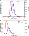
SEC-MALS of S100B and p53-(293-393) oligomers. A, 250 μm dimer p53-(293-393)-L344A (blue) and the complex of 200 μm p53 variant and 100 μm S100B (red) differed in tr and formula imagew determined by MALS. B, the elution profile of 300 μm p53-(293-393) and the complex of 300 μm p53 with 150 μm S100B differed in tr and formula imagew. No additional peaks of S100B in complex with lower oligomeric forms of p53 were detected. WT, wild type.
FIGURE 4.
FIGURE 4.
Analytical AUC of p53 QMFL-FlAsH with S100 proteins. 250 nm of p53-QMFL-FlAsH and different amounts of S100 were run at 45,000 rpm. The sedimentation profile was fitted to a double Gaussian equation, and the Mr was calculated with SedFit.
FIGURE 5.
FIGURE 5.
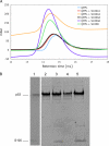
SEC-PAGE of p53-QMFL and S100. A, the analytical gel filtrations of p53-QMFL in complex with different S100 proteins (in a ratio of 1 to 2) resulted in small tr shifts for S100B, S100A2, and S100A6. mAU, milliabsorbance units. B, the elution between 14 and 15 ml was collected, concentrated, and analyzed via SDS-PAGE. Lane 1, QMFL + S100B; lane 2, QMFL + S100A1; lane 3, QMFL + S100A2; lane 4, QMFL + S100A4; lane 5, QMFL + S100A6.
FIGURE 6.
FIGURE 6.
Binding of S100 proteins to the transactivation domain of p53. S100 proteins were titrated to 0. 5 μm p53-(1-57)-Lys-methoxycoumarin in fluorescence anisotropy experiments.
FIGURE 7.
FIGURE 7.
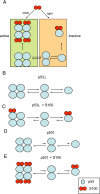
Binding model of S100 and p53. A, proteins of the S100 family can bind p53 as a tetramer as well as a monomer. The different oligomeric forms of p53 are in equilibrium. S100 can have an activating function binding to the tetramer or an inhibiting effect binding to the monomer of p53. B, at low concentrations of p53 (below the Kd for tetramerization), there is a significant equilibrium between tetramer, dimer, and monomer of p53. C, the tight binding of S100 to the monomer will displace the oligomerization equilibrium (illustrated by the different lengths of the equilibrium arrows) in favor of the monomer and consequently inhibit p53 function. D, when the concentration of p53 is much higher than the Kd for tetramerization, almost all of p53 is present as a tetramer. E, under these circumstances, the tight binding of S100 to the p53 monomer will not significantly alter the tetramerization equilibrium, and the inhibiting effect is overwhelmed by the activating or stabilizing effect of S100 binding to the tetramer of p53.

References

    1. Donato, R. (2003) Microsc. Res. Tech. 60 540-551 - PubMed
    1. Moore, B. W. (1965) Biochem. Biophys. Res. Commun. 19 739-744 - PubMed
    1. Koch, M., Bhattacharya, S., Kehl, T., Gimona, M., Vasak, M., Chazin, W., Heizmann, C. W., Kroneck, P. M., and Fritz, G. (2007) Biochim. Biophys. Acta 1773 457-470 - PubMed
    1. Ostendorp, T., Leclerc, E., Galichet, A., Koch, M., Demling, N., Weigle, B., Heizmann, C. W., Kroneck, P. M., and Fritz, G. (2007) EMBO J. 26 3868-3878 - PMC - PubMed
    1. Donato, R. (2001) Int. J. Biochem. Cell Biol. 33 637-668 - PubMed

MeSH terms