. 2009 May 15;284(20):13589-13601.
doi: 10.1074/jbc.M809244200. Epub 2009 Mar 18.

Hepatitis C virus NS5A protein is a substrate for the peptidyl-prolyl cis/trans isomerase activity of cyclophilins A and B

Affiliations

Hepatitis C virus NS5A protein is a substrate for the peptidyl-prolyl cis/trans isomerase activity of cyclophilins A and B

Xavier Hanoulle et al. J Biol Chem. .

Abstract

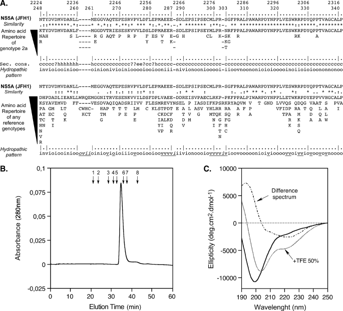
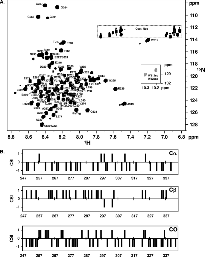
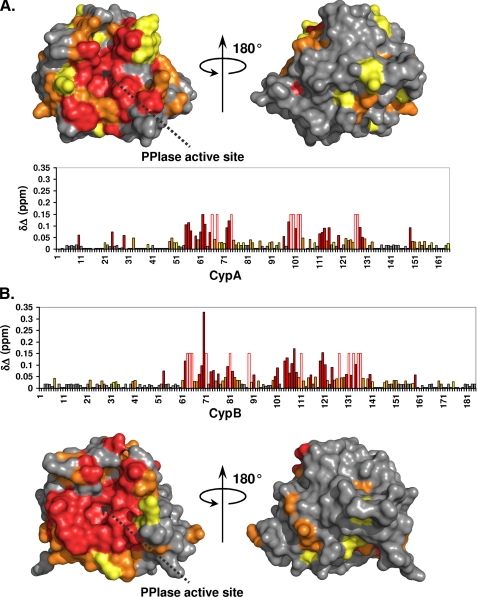
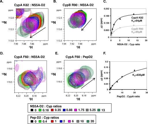
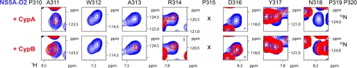
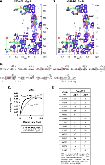
We report here a biochemical and structural characterization of domain 2 of the nonstructural 5A protein (NS5A) from the JFH1 Hepatitis C virus strain and its interactions with cyclophilins A and B (CypA and CypB). Gel filtration chromatography, circular dichroism spectroscopy, and finally NMR spectroscopy all indicate the natively unfolded nature of this NS5A-D2 domain. Because mutations in this domain have been linked to cyclosporin A resistance, we used NMR spectroscopy to investigate potential interactions between NS5A-D2 and cellular CypA and CypB. We observed a direct molecular interaction between NS5A-D2 and both cyclophilins. The interaction surface on the cyclophilins corresponds to their active site, whereas on NS5A-D2, it proved to be distributed over the many proline residues of the domain. NMR heteronuclear exchange spectroscopy yielded direct evidence that many proline residues in NS5A-D2 form a valid substrate for the enzymatic peptidyl-prolyl cis/trans isomerase (PPIase) activity of CypA and CypB.


Figures

FIGURE 1.
FIGURE 1.
Sequence analysis and biochemical characterization of NS5A-D2 domain 2 from HCV. A, amino acid repertoires. The NS5A-D2 aa 248–341 sequence from the HCV JFH1 strain of genotype 2a (GenBank™ accession number AB047639), which was used in this study, is indicated. Amino acids are numbered with respect to NS5A and the HCV JFH1 polyprotein (top row). The hyphens indicate the aa deletions compared with the sequence alignment of any genotypes (see below). The aa repertoire deduced from the ClustalW multiple alignments of 21 NS5A sequences of genotype 2a is shown at the top. Amino acids observed at a given position less than twice were not included. At the bottom is the aa repertoire of the 27 representative NS5A sequences from confirmed HCV genotypes and subtypes (listed with accession numbers in Table 1 in Ref. ; see the European HCV Database for details). The degree of aa and physicochemical conservation at each position can be inferred from the extent of variability (with the observed aa listed in decreasing order of frequency from top to bottom) together with the similarity index according to ClustalW convention (asterisk, invariant; colon, highly similar; dot, similar (40)) and the consensus hydropathic pattern deduced from the consensus aa repertoire: o, hydrophobic position (Phe, Ile, Trp, Tyr, Leu, Val, Met, Pro, Cys); n, neutral position (Gly, Ala, Thr, Ser); i, hydrophilic position (Lys, Gln, Asn, His, Glu, Asp, Arg); v, variable position (i.e. when both hydrophobic and hydrophilic residues are observed at a given position). A position that is underlined in the hydropathic pattern for any reference genotypes (bottom) indicates the change of position status when compared with the hydropathic pattern for genotype 2a (middle). Secondary structure predictions of NS5A-D2 are indicated as helical (h), extended (e), undetermined (coil (c)), or ambiguous (?). Sec. Cons., consensus of protein secondary structures predictions for NS5A-D2 from JFH1 strain deduced from a large set of prediction methods available at the NPSA website, including DSC, HNNC, MLRC, PHD, Predator, SOPM, and SIMPA96 available at the NPSA website (see Ref. and references therein). B, gel filtration analysis of NS5A-D2 was performed on a Superdex S200 column equilibrated in 50 mm sodium phosphate, pH 7.4, 1 mm Tris(2-carboxyethyl) phosphine hydrochloride with a flow rate of 0.5 ml/min. Elution volumes of globular protein standards are indicated by black arrows with the following corresponding molecular masses: 1, thyroglobulin (669,000 Da); 2, ferritin (44,0000 Da); 3, aldolase (158,000 Da); 4, conalbumin (75,000 Da); 5, ovalbumin (43,000 Da); 6, chymotrypsin (25,000 Da); 7, ribonuclease (13,700 Da); 8, vitamin B12 (1,355 Da). C, Far-UV circular dichroism analysis of 8 μm NS5A-D2 in 10 mm sodium phosphate, pH 7.4, 1 mm Tris(2-carboxyethyl) phosphine hydrochloride (solid line) complemented with 50% TFE (dotted line). The difference spectrum (alternating dashed line) was obtained by subtracting the latter spectrum from the former.
FIGURE 2.
FIGURE 2.
NMR characterization of NS5A-D2 (JFH1). A, assigned 1H,15N HSQC spectrum of domain 2 of NS5A (JFH1). The smaller insert shows the spectrum region corresponding to the two tryptophan side chains. B, CSI analysis of NS5A-D2. NS5A-D2 13Cα, 13Cβ, and 13CO chemical shifts were analyzed using CSI software (54). The consensus CSI is zero along the complete sequence and therefore is not shown.
FIGURE 3.
FIGURE 3.
Cyclophilin binding sites for NS5A-D2. The 1H and 15N combined chemical shift perturbations (δΔ) induced on the CypA (A) or CypB (B) spectra following NS5A-D2 addition in a 1:1 molar ratio were plotted along the cyclophilin primary sequences and on their respective three-dimensional molecular surfaces. Residues with combined chemical shift perturbations 0.02 ≤ δΔ ≤ 0.03 ppm are in yellow; 0.03 ≤ δΔ ≤ 0.05 ppm are in orange; and δΔ> 0.05 ppm are in red. For cyclophilin residues for which the proton amide resonances disappear due to important line broadening in the presence of NS5A-D2, a fixed δΔ value of 0.15 ppm was set. These residues are depicted by an open bar circled in red in the diagrams. The PPIase active site of cyclophilins is indicated by a dotted black arrow.
FIGURE 4.
FIGURE 4.
Titration experiments between cyclophilins and NS5A-D2 or PepD2. Panels A, B, D, and E correspond to the superimposition of the 1H,15N HSQC spectra of CypA (or CypB, in B) acquired in the presence of increasing amounts of unlabeled NS5A-D2 (A, B, and D) or Pep-D2 (E) (PepD2 corresponds to residues 304–323 of NS5A-D2: 304GFPRALPAWARPDYNPPLVE323). Lys82 in CypA is equivalent to Arg90 in CypB and is at the periphery of the NS5A-D2 binding site. C, titration curves corresponding to experiments in A (black triangle) and B (gray diamonds). The 1H,15N combined chemical shift perturbations Δδ (in ppm) (Δδ = (δ(1H)2 + 0.2.δ(15N)2)½) were plotted as a function of the NS5A-D2:cyclophilin molar ratios. The dissociation constants (KD) were obtained by fitting the experimental data with the following equation: KD = [Cypfree]·[NS5A-D2free]/[Cyp:NS5A-D2]. D and E, Phe60 in CypA is directly in the binding site of NS5A-D2 and broadens when titrated with the D2 domain (D) but not with Pep-D2 (E). F, titration curve corresponding to experiments in E (black triangles).
FIGURE 5.
FIGURE 5.
A major interaction site of NS5A-D2 with CypA and CypB. Each panel corresponds to the superposition of a 1H,15N HSQC spectrum acquired on NS5A-D2 alone (in blue) and of a 1H,15N plane obtained with the HNnoCO pulse sequence that specifically selects the NS5A-D2 subspectrum in a NS5A-D2/Cyp (1:1) sample (in red). The motif 310PAWARPDYNPP320 of NS5A-D2 interacts with CypA.
FIGURE 6.
FIGURE 6.
Cis/trans isomerization of HCV NS5A-D2 X-Pro peptide bonds catalyzed by CypA and CypB. 1H,15N heteronuclear exchange spectra recorded at 800 MHz with mixing times of 0.88 ms (in blue) and 100 ms (in red) on [15N]NS5A-D2 samples (220 μm) with catalytic amounts of either CypA (A) or CypB (B) (23 μm). The NMR resonances (trans, cis, and the two exchange peaks) of NS5A-D2 residues for which the PPIase activity of a cyclophilin can be evidenced are connected by green dotted boxes. C, amino acid sequence of NS5A-D2 (JFH1) construction. Residues on which PPIase activities of CypA and CypB have been monitored are bold and shown in violet. The 15 proline residues of NS5A-D2 are indicated on a light gray background. The previously defined binding site of cyclophilins is boxed, and residues that are affected only by CypA binding (see Fig. 4 and under “Results”) are marked with asterisks. D, determination of the CypA (in black (Δ)) and CypB-catalyzed (in gray (×)) exchange rates toward the Val274-Pro275 NS5A-D2 peptide bond. Experimental data measured on Val274 for (Itc/Itt) (CypA (Δ); CypB (×)) were fitted to the theoretical Equation 2 (see “Experimental Procedures”) (CypA, black line; CypB, gray line). E, resulting exchange rates (kexch) of the cis/trans isomerization processes in NS5A-D2 catalyzed by the addition of catalytic amounts of either CypA or CypB.

Similar articles

Cited by

References

    1. National Institutes of Health (2002) Hepatology 36 Suppl. 1, S2-S20
    1. Appel, N., Schaller, T., Penin, F., and Bartenschlager, R. (2006) J. Biol. Chem. 281 9833-9836 - PubMed
    1. Moradpour, D., Penin, F., and Rice, C. M. (2007) Nat. Rev. 5 453-463 - PubMed
    1. Tellinghuisen, T. L., Evans, M. J., von Hahn, T., You, S., and Rice, C. M. (2007) J. Virol. 81 8853-8867 - PMC - PubMed
    1. Lohmann, V., Korner, F., Koch, J., Herian, U., Theilmann, L., and Bartenschlager, R. (1999) Science 285 110-113 - PubMed

Publication types

MeSH terms

LinkOut - more resources