. 2009 Jun 12;284(24):16659-16666.
doi: 10.1074/jbc.M808029200. Epub 2009 Mar 18.

Structural insight into the activation mechanism of human pancreatic prophospholipase A2

Affiliations

Structural insight into the activation mechanism of human pancreatic prophospholipase A2

Wei Xu et al. J Biol Chem. .

Abstract

Pancreatic phospholipase A2 (phospholipase A2 group 1B, G1B) belongs to the superfamily of secreted phospholipase A2 (PLA2) enzymes. G1B has been proposed to be a potential target for diseases such as hypertension, obesity, and diabetes. Human pancreatic prophospholipase A2 (pro-hG1B) is activated by cleavage of the first seven-residue propeptide (phospholipase A2 propeptide, PROP). However, questions still remain on the mode of action for pro-hG1B. In this work, we expressed pro-hG1B in Pichia pastoris and determined the crystal structure at 1.55-A resolution. The x-ray structure demonstrates that pro-hG1B forms a trimer. In addition, PROP occupies the catalytic cavity and can be self-cleaved at 37 degrees C. A new membrane-bound surface and activation mechanism are proposed based on the trimeric model of pro-hG1B. We also propose a new autoproteolytic mechanism for pro-hG1B by the reaction triad Asp49-Arg0-Ser(-2) that is similar to the serine protease catalytic triad.


Figures
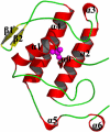
FIGURE 1.
FIGURE 1.
Structure of pro-hG1B. The overall structure of human pancreatic prophospholipase A2 is shown. Helices are in red, and β-strands are in yellow. Loops are colored in green. The sulfate ion is presented as a CPK model in magenta.
FIGURE 2.
FIGURE 2.
Arg0 occupies the active site. Stereo view of the catalytic site with 2Fo-Fc electron density map contoured at 1σ. Arg0 interacts with Tyr22, Gly30, His48, and Asp49 through hydrogen bonds.
FIGURE 3.
FIGURE 3.
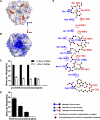
The trimeric form of pro-hG1B. A, electrostatic surface potential representation of pro-hG1B with the PROP occupying the active site cavity. The PROP is shown as a scheme and is colored in green. The surface potential is calculated using ABPS (47) and color-coded, where red and blue represent net negative and positive charges, and white represents overall neutral positions, respectively. The total charge is represented from −8 kbT/ec to 8 kbT/ec as the color change, in which kb is the Boltzmann constant, T is the temperature, and ec is the electron charge. B, view of molecule with 180 degree rotation around the y axis of A. The sulfate ion is shown as a magenta CPK model. C, calculated molecular weight of pro-hG1B obtained from DLS experiments in various protein concentrations, 30, 15, 7.5, 3.7, and 0 mg/ml, respectively, and with/without lithium sulfate. All data shown are the average of triplicates. Error bars denote the S.E. D, calculated molecular weight of hG1B obtained from DLS experiments in various protein concentrations, 30, 7.5, 3.7, and 0 mg/ml, respectively, with lithium sulfate. All data shown are the average of triplicates. Error bars denote the S.E. Panels C and D were prepared by GraphPad Prism 5. E, subunit interface of pro-hG1B trimer. Two monomer chains, chain B and chain C, are colored in blue and red, respectively. The key residues involved in the subunit interaction are labeled. The figure was prepared by LIGPLOT (48).
FIGURE 4.
FIGURE 4.
Autocleavage of PROP. A, mass spectrometry data of pro-hG1B (100 μg) digested with 2 μg of trypsin or thrombin, pro-hG1B at 37 °C for 18 h in 100 μl of 10 mm Tris, pH 8. 5. Pro-hG1B stored at 4 °C was used as a blank control. B, purified pro-hG1B and hG1B (activated by thrombin) were dissolved in 10 μl of 10 mm Tris, pH 8.0, 150 mm NaCl, then added to an equal volume of SDS-PAGE loading buffer that contained 2% (w/v) SDS and heated for 15 min when marked with +. C, hG1B (activated by autocleavage) was dissolved in 10 μl of 10 mm Tris, pH 8.0, 150 mm NaCl, then added to an equal volume of SDS-PAGE loading buffer that contained 2% (w/v) SDS and heated for 15 min at the indicated temperature.
FIGURE 5.
FIGURE 5.
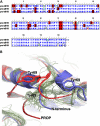
69-loop adopts a unique conformation. A, sequence alignment between human pro-G1B and pro-G1B from porcine and bovine, color-coded as following: blue for identical alignment, red shading for semi-conserved substitution, and other colors for no similarity. The alignment was performed using BIOEDIT (49). B, superposition of all published pancreatic PLA2 in the 69-loop region. Pro-hG1B is colored in red, pro-pG1B (PDB code: 1HN4) is colored in blue, others (porcine and bovine G1B) are colored in green. Tyr69 is presented as a stick model.
FIGURE 6.
FIGURE 6.
Asp49-Arg0-Ser(-2) reaction triad. Pair fitting of the proposed pro-hG1B triad with the trypsin catalytic triad is shown. Asp49, Ser-2, and Arg0 of pro-hG1B are colored in orange. Asp102, His57, and Ser95 of trypsin (PDB code: 1K1I) are colored in cyan.

References

    1. Schaloske R. H., Dennis E. A. ( 2006) Biochim. Biophys. Acta 1761, 1246– 1259 - PubMed
    1. Frossard P. M., Lestringant G. G. ( 1995) Clin. Genet. 48, 284– 287 - PubMed
    1. Kuopio T., Ekfors T. O., Nikkanen V., Nevalainen T. J. ( 1995) Apmis. 103, 69– 78 - PubMed
    1. Huggins K. W., Boileau A. C., Hui D. Y. ( 2002) Am. J. Physiol. Endocrinol. Metab. 283, E994– 1001 - PubMed
    1. Labonte E. D., Kirby R. J., Schildmeyer N. M., Cannon A. M., Huggins K. W., Hui D. Y. ( 2006) Diabetes 55, 935– 941 - PMC - PubMed

Publication types

Substances