. 2009 May 15;284(20):13296-13300.
doi: 10.1074/jbc.C800241200. Epub 2009 Mar 17.

FoxO1 controls insulin-dependent adipose triglyceride lipase (ATGL) expression and lipolysis in adipocytes

Affiliations

FoxO1 controls insulin-dependent adipose triglyceride lipase (ATGL) expression and lipolysis in adipocytes

Partha Chakrabarti et al. J Biol Chem. .

Abstract

FoxO1 represents a central regulator of metabolism in several cell types. Although FoxO1 is abundant in adipocytes, its biological functions in these cells remain largely unknown. We show here that the promotor region of the rate-limiting lipolytic enzyme, adipose triglyceride lipase (ATGL), has two FoxO1-binding sites, and co-transfection with wild type and unphosphorylated FoxO1 mutant activates the expression of luciferase driven by the ATGL promotor. In 3T3-L1 adipocytes, insulin controls nucleo-cytoplasmic shuttling of FoxO1 and regulates its interaction with endogenous ATGL promotors. Knockdown of FoxO1 in 3T3-L1 adipocytes decreases the expression of ATGL and attenuates basal and isoproterenol-stimulated lipolysis. Infection of mouse embryonic fibroblasts with FoxO1-encoding lentivirus increases ATGL expression and renders it sensitive to regulation by insulin. Thus, FoxO1 may play an important role in the regulation of lipolysis in adipocytes by controlling the expression of ATGL.


Figures

FIGURE 1.
FIGURE 1.
ATGL is a transcriptional target of FoxO1. A, 3T3-L1 adipocytes were differentiated for the indicated time periods, and FoxO1 and ATGL protein levels were detected in whole cell lysates (50 μg). CEBPα was used as positive control for differentiation and cellugyrin as loading control. B, 293T cells were transiently transfected with 2979/+21 luciferase (LUC) ATGL promoter construct together with empty vector (EV), FoxO1, or FoxO1-AAA constructs. For normalization of transfection efficiency, cells were co-transfected with eGFP. After 48 h, cells were harvested in the reporter lysis buffer. Luciferase activity in cell lysates was assayed as described under “Experimental Procedures” and normalized by GFP fluorescence. Data are presented for triplicate samples as mean ± S.D. (error bars). **, p < 0.001. C, differentiated 3T3-L1 cells were left in complete medium (Control), incubated in DMEM without serum (Starve), or treated with 100 nm insulin (Ins) for 16 h. Fixed cells were stained with FoxO1 antibody and Alexa Fluor 488-conjugated donkey anti-rabbit IgG (red). 4′,6-Diamidino-2-phenylindole (DAPI) (blue) was incorporated in the mounting solution. D, 3T3-L1 adipocytes were treated as indicated for panel C, and nuclear and cytosolic fractions were isolated and analyzed by Western blotting (50 μg per lane). Glyceraldehyde-3-phosphate dehydrogenase (GAPDH) and RNA polymerase II (Pol II) were used as controls for cytosolic and nuclear fractions, respectively. E, ChIP assays were performed in 3T3-L1 adipocytes incubated either in complete medium (Serum +) or in DMEM without serum (Serum –) for 16 h. Following cross-linking and sonication, genomic fragments were immunoprecipitated with antibody against FoxO1 or rabbit IgG, and then 221 bp between nucleotides –1004 and –1225 of the ATGL promotor was amplified by PCR, separated in a 3% agarose gel, and visualized by ethidium bromide staining. F, 3T3-L1 cells were incubated in DMEM (Starve) or DMEM with 100 nm Ins for 16 h. The region between nucleotides –1041 and –1158 of the ATGL promotor in the immunoprecipitated genomic fragments was amplified by SYBR green reaction as described under “Experimental Procedures” and analyzed by quantitative PCR. The experiment was performed in triplicate, and data were expressed as mean ± S.D. (error bars) normalized by input. *, p < 0.05.
FIGURE 2.
FIGURE 2.
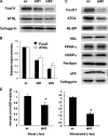
Knockdown of FoxO1 with siRNA decreases lipolysis and ATGL expression in adipocytes. A, differentiated 3T3-L1 adipocytes (day 7) were transfected with either 30 nm scrambled (sc) or two different siRNAs against mouse FoxO1 (si#1 and si#2), and total cell lysates were harvested after 48 h. Levels of ATGL protein upon knockdown of FoxO1 were analyzed by Western blotting. B, quantitative analysis of relative band intensities of FoxO1 and ATGL in three independent experiments as described for panel A. Data were presented as mean ± S.D. (error bars). C, analysis of adipocyte differentiation markers and proteins associated with lipolysis upon knockdown of FoxO1. D, glycerol release in the media was determined in cells transfected with either scrambled (sc) or specific (si#2) siRNA for 2 h. Data were expressed as mean ± S.D. (error bars) relative to non-stimulated scrambled cells. Iso, isoproterenol. All panels, *, p < 0.05.
FIGURE 3.
FIGURE 3.
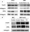
FoxO1 confers insulin sensitivity to regulation of ATGL expression in mouse embryonic fibroblasts. A, Western blot analysis of FoxO1 and ATGL expression in total cell lysates prepared from 3T3-L1 adipocytes (left panels) and MEFs (right panels) incubated either in complete medium (Control) or in DMEM in the absence (Starve) or presence of 100 nm Ins for 16 h. B, MEFs stably overexpressing FoxO1 (MEF-FoxO1) were treated as described for panel A, and total cell lysates were analyzed by Western blotting. Wild-type MEF was shown as control.

References

    1. Fraze, E., Donner, C. C., Swislocki, A. L., Chiou, Y. A., Chen, Y. D., and Reaven, G. M. (1985) J. Clin. Endocrinol. Metab. 61 807–811 - PubMed
    1. Stumvoll, M., Goldstein, B. J., and van Haeften, T. W. (2008) Lancet 371 2153–2156 - PubMed
    1. Wymann, M. P., and Schneiter, R. (2008) Nat. Rev. Mol. Cell Biol. 9 162–176 - PubMed
    1. Zechner, R., Kienesberger, P. C., Haemmerle, G., Zimmermann, R., and Lass, A. (2009) J. Lipid Res. 50 3–21 - PubMed
    1. Duncan, R. E., Ahmadian, M., Jaworski, K., Sarkadi-Nagy, E., and Sul, H. S. (2007) Annu. Rev. Nutr. 27 79–101 - PMC - PubMed

Publication types

MeSH terms