. 2009 Jun;83(11):5683-92.
doi: 10.1128/JVI.00231-09. Epub 2009 Mar 18.

Type I and type II interferons inhibit the translation of murine norovirus proteins

Affiliations

Type I and type II interferons inhibit the translation of murine norovirus proteins

Harish Changotra et al. J Virol. 2009 Jun.

Abstract

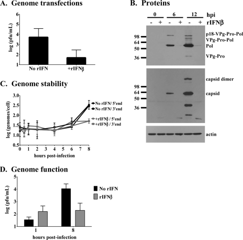
Human noroviruses are responsible for more than 95% of nonbacterial epidemic gastroenteritis worldwide. Both onset and resolution of disease symptoms are rapid, suggesting that components of the innate immune response are critical in norovirus control. While the study of the human noroviruses has been hampered by the lack of small animal and tissue culture systems, our recent discovery of a murine norovirus (MNV) and its in vitro propagation have allowed us to begin addressing norovirus replication strategies and immune responses to norovirus infection. We have previously demonstrated that interferon responses are critical to control MNV-1 infection in vivo and to directly inhibit viral replication in vitro. We now extend these studies to define the molecular basis for interferon-mediated inhibition. Viral replication intermediates were not detected in permissive cells pretreated with type I interferon after either infection or transfection of virion-associated RNA, demonstrating a very early block to virion production that is after virus entry and uncoating. A similar absence of viral replication intermediates was observed in infected primary macrophages and dendritic cells pretreated with type I IFN. This was not due to degradation of incoming genomes in interferon-pretreated cells since similar levels of genomes were present in untreated and pretreated cells through 6 h of infection, and these genomes retained their integrity. Surprisingly, this block to the translation of viral proteins was not dependent on the well-characterized interferon-induced antiviral molecule PKR. Similar results were observed in cells pretreated with type II interferon, except that the inhibition of viral translation was dependent on PKR. Thus, both type I and type II interferon signaling inhibit norovirus translation in permissive myeloid cells, but they display distinct dependence on PKR for this inhibition.


Figures

FIG. 1.
FIG. 1.
Type I IFN potently inhibits MNV-1 replication in Mφs and DCs. (A) Different IFN treatment conditions (pretreatment or posttreatment with respect to infection) were tested for MNV-1.CW1 inhibition. For pretreatment, RAW264.7 cells were pretreated with 1,000 U of rIFN-β/ml for 18 to 20 h prior to infection. For posttreatment, RAW264.7 cells were treated with 100 U of rIFN-β/ml immediately after infection. Cells were infected with MNV-1.CW1 at an MOI of 1 for 1 h on ice, supernatants were collected 24 hpi, and virus titers determined by plaque assay. (B) RAW264.7 cells were untreated or pretreated with increasing amounts of rIFN-β for 18 to 20 h. Cells were then infected with MNV-1.CW1 at an MOI of 0.1, 1, or 10 for 1 h on ice. Supernatants were collected 24 hpi, and the virus titers were determined by plaque assay. (C) WBC264-9C, J774.1, P388D1, and IC-21 Mφ cell lines were pretreated with various amounts of rIFN-β for 18 to 20 h prior to infection with MNV-1.CW1 at an MOI of 1, based on titration of the virus stock on RAW264.7 cells. Titers of supernatants collected 24 hpi were determined by plaque assay. The data are presented as the average fold increases in inhibition comparing the number of virions in rIFNβ-pretreated cells to the number in untreated cells, on a logarithmic scale. (D) Primary BMMφs and BMDCs were differentiated from 129SvEv mice. Cells were treated with increasing amounts of rIFN-β for 18 to 20 h prior to infection with MNV-1.CW1 at an MOI of 1. Titers of supernatants collected 24 hpi were determined by plaque assay. In all cases, duplicate wells per condition were tested in individual experiments, and data from three or more independent experiments are averaged.
FIG. 2.
FIG. 2.
Type I IFN inhibits early MNV-1 translation. In all experiments, RAW264.7 cells were either untreated or pretreated with 1,000 U of rIFN-β/ml for 18 to 20 h. (A) Cells were then transfected with 0.2 μg of virion-associated RNA. Supernatants were collected 24 h posttransfection, and virus titers were determined by plaque assay. Duplicate wells per condition were tested in three independent experiments, and the data were averaged. The limit of detection is 10 PFU/ml. Infectious virions were only detected in three of the six supernatants tested from rIFN-β-pretreated cells. (B) Cells were infected with MNV-1.CW1 at an MOI of 1 for 1 h on ice. Cell lysates were collected at 0, 6, or 12 hpi and analyzed by immunoblotting. Blots were first probed with α-ProPol, then stripped and reprobed with α-capsid, and finally stripped and reprobed with α-actin antibody as a loading control. The data are representative of three experiments, with duplicate wells tested in each experiment. (C) Cells were infected with MNV-1.CW1 at an MOI of 10, or mock infected, for 1 h on ice. Total RNA was extracted from cell lysates at 0, 0.5, 1, 2, 4, 6, or 8 hpi; single-stranded cDNA was generated; and triplicate qRT-PCRs were performed with primers specific to the 5′ end of the MNV-1 genome, the 3′ end of the MNV-1 genome, or 18S rRNA for normalization. No virus signal was detected in mock-infected cultures (data not shown). The data from two experiments were averaged, with duplicate wells tested in each experiment. (D) Cells were infected with MNV-1.CW1 at an MOI of 10 for 1 h on ice. Total RNA was extracted from untreated or rIFN-β-pretreated cells at 1 or 8 hpi, and 2 μg was transfected into 2 × 10⁵ untreated cells using Lipofectamine 2000. Supernatants were collected at 24 h posttransfection, and virus titers were determined by plaque assay. Duplicate wells per condition were tested in two independent experiments, and the data were averaged. The limit of detection is 20 PFU/ml.
FIG. 3.
FIG. 3.
Type II IFN inhibits early MNV-1 translation. (A and B) RAW264.7 cells were untreated or pretreated with increasing amounts of rIFN-γ for 18 to 20 h. Cells were then infected with MNV-1.CW1 at an MOI of 1 for 1 h on ice (A) or transfected with 0.2 μg of virion-associated RNA (B). Supernatants were collected 24 h later, and virus titers were determined by plaque assay. Duplicate wells per condition were tested in two independent experiments, and the data were averaged. The limit of detection was 10 PFU/ml. (C to E) RAW264.7 cells were either untreated or pretreated with 1,000 U of rIFN-γ/ml for 18 to 20 h. (C) Cells were infected with MNV-1.CW1 at an MOI of 1 for 1 h on ice. Cell lysates were collected at 0, 6, or 12 hpi and analyzed by immunoblotting. Blots were first probed with α-ProPol, then stripped and reprobed with α-capsid, and finally stripped and reprobed with α-actin antibody as a loading control. The data are representative of two experiments, with duplicate wells tested in each experiment. (D) Cells were infected with MNV-1.CW1 at an MOI of 10, or mock infected, for 1 h on ice. Total RNA was extracted from cell lysates at 0, 0.5, 1, 2, 3, 4, 6, or 8 hpi; single-stranded cDNA was generated, and triplicate qRT-PCRs were performed with primers specific to the 5′ end of the MNV-1 genome or 18S rRNA for normalization. No virus signal was detected in mock-infected cultures (data not shown). The data from two experiments were averaged, with duplicate wells tested in each experiment. (D) Cells were infected with MNV-1.CW1 at an MOI of 10 for 1 h on ice. Total RNA was extracted from untreated or rIFN-γ-pretreated cells at 1 or 8 hpi, and 2 μg was transfected into 2 × 10⁵ untreated cells using Lipofectamine 2000. Supernatants were collected 24 h posttransfection, and virus titers were determined by plaque assay. Duplicate wells per condition were tested in two independent experiments, and the data were averaged. The limit of detection is 20 PFU/ml.
FIG. 4.
FIG. 4.
Type I and type II IFN inhibit MNV-1 translation in primary DCs. BMDCs were differentiated from wild-type 129SvEv (WT) mice (A and B) or triply deficient PKR/RNaseL/Mx1−/− (TD) mice (C and D) and then either left untreated or pretreated with 1,000 U of rIFN-β (A and C) or rIFN-γ (B and D)/ml for 18 to 20 h. Cells were then mock infected or infected with MNV-1.CW1 at an MOI of 1 for 1 h on ice. Cell lysates were collected at 0, 6, 12, and 24 hpi for MNV-1.CW1-infected cultures and at 24 hpi for mock-infected cultures (24M) and analyzed by immunoblotting with antibody to ProPol. Blots were stripped and reprobed with α-actin antibody as a loading control. The data are representative of three experiments. (E) WT or TD BMDCs were either left untreated, pretreated with 1,000 U of rIFN-β/ml, or pretreated with 1,000 U of rIFN-γ/ml for 18 to 20 h. Cells were infected with MNV-1.CW1 at an MOI of 1 for 1 h on ice. Supernatants were collected at 0, 6, 9, and 12 hpi, and virus titers were determined by plaque assay. Duplicate wells per condition were tested, and the data were averaged. The number of virions at each time point after 0 hpi was then normalized to the number of virions detected at 0 hpi, which was assumed to be the level of input virus. The experiment was repeated twice, and the data were averaged.
FIG. 5.
FIG. 5.
Type I, but not type II, IFN inhibits a late step in the MNV-1 replication cycle. WT (A) or TD (B) BMDCs were either left untreated, pretreated with 1,000 U of rIFN-β/ml, or pretreated with 1,000 U of rIFN-γ/ml for 18 to 20 h. Cells were infected with MNV-1.CW1 at an MOI of 1 for 1 h on ice. Supernatants were collected at 18, 24, 48, and 72 hpi, and virus titers were determined by plaque assay. Duplicate wells per condition were tested in two independent experiments, and the data were averaged. The limit of detection is 10 PFU/ml. Virus titers from earlier time points (0, 6, 9, and 12 hpi) are included from Fig. 4 for clarity. (C) The same data presented in panels A and B are replicated to highlight the differences observed in WT and TD cells.

Similar articles

Cited by

References

    1. Burroughs, J. N., and F. Brown. 1978. Presence of a covalently linked protein on calicivirus RNA. J. Gen. Virol. 41443-446. - PubMed
    1. Centers for Disease Control and Prevention. 2005. Infectious disease and dermatologic conditions in evacuees and rescue workers after Hurricane Katrina-multiple states, August-September, 2005. MMWR Morb. Mortal. Wkly. Rep. 54961-964. - PubMed
    1. Centers for Disease Control and Prevention. 2005. Norovirus outbreak among evacuees from hurricane Katrina-Houston, Texas, September 2005. MMWR Morb. Mortal. Wkly. Rep. 541016-1018. - PubMed
    1. Centers for Disease Control and Prevention. 2002. Outbreak of acute gastroenteritis associated with Norwalk-like viruses among British military personnel-Afghanistan, May 2002. MMWR Morb. Mortal. Wkly. Rep. 51477-479. - PubMed
    1. Chang, K. O., and D. W. George. 2007. Interferons and ribavirin effectively inhibit Norwalk virus replication in replicon-bearing cells. J. Virol. 8112111-12118. - PMC - PubMed

Publication types