. 2009 Jun;83(11):5735-48.
doi: 10.1128/JVI.02440-08. Epub 2009 Mar 18.

Cell-to-cell spread of the RNA interference response suppresses Semliki Forest virus (SFV) infection of mosquito cell cultures and cannot be antagonized by SFV

Affiliations

Cell-to-cell spread of the RNA interference response suppresses Semliki Forest virus (SFV) infection of mosquito cell cultures and cannot be antagonized by SFV

Ghassem Attarzadeh-Yazdi et al. J Virol. 2009 Jun.

Abstract

In their vertebrate hosts, arboviruses such as Semliki Forest virus (SFV) (Togaviridae) generally counteract innate defenses and trigger cell death. In contrast, in mosquito cells, following an early phase of efficient virus production, a persistent infection with low levels of virus production is established. Whether arboviruses counteract RNA interference (RNAi), which provides an important antiviral defense system in mosquitoes, is an important question. Here we show that in Aedes albopictus-derived mosquito cells, SFV cannot prevent the establishment of an antiviral RNAi response or prevent the spread of protective antiviral double-stranded RNA/small interfering RNA (siRNA) from cell to cell, which can inhibit the replication of incoming virus. The expression of tombusvirus siRNA-binding protein p19 by SFV strongly enhanced virus spread between cultured cells rather than virus replication in initially infected cells. Our results indicate that the spread of the RNAi signal contributes to limiting virus dissemination.


Figures
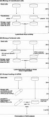
FIG. 1.
FIG. 1.
Flow charts for cell mixing and scrape-loading experiments involving U4.4 mosquito cells. (A) Mixing of transfected cells. (B) Mixing of infected cells. (C) Scrape loading of fluorescein-labeled siRNA into cells. Experimental details such as cell numbers, etc., are described in Materials and Methods.
FIG. 2.
FIG. 2.
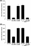
SFV interactions with RNAi. (A) U4.4 mosquito cells were transfected with pRL-CMV (which expresses RLuc) (20 ng) and RLuc or negative control (nc) siRNAs (5 nM) for 5 h and infected with SFV4 (MOI of 10) at 24 h posttransfection, and luciferase activities were measured at 24 h p.i. (B) U4.4 mosquito cells were transfected with pRL-CMV (20 ng), infected with SFV4 (MOI of 10) at 24 h posttransfection, and then transfected with RLuc or negative control siRNAs (5 nM) for 5 h, and luciferase activities were measured 24 h posttransfection. Each bar represents the mean of three replicates; error bars indicate standard deviations. Every experiment was repeated at least twice.
FIG. 3.
FIG. 3.
SFV interactions with antiviral RNAi. (A) SFV4(3H)-RLuc encodes RLuc (flanked by duplicated nsP2-protease cleavage sites at the nsP3/4 junction) as part of the viral nonstructural polyprotein. (B) Transfection of RLuc but not negative control (nc) siRNA (10 nM) of U4.4 mosquito cells with SFV4(3H)-RLuc (MOI of 10) at 1 or 8 h p.i. strongly reduced virus replication. RLuc activities were determined at 24 h posttransfection. NTC, nontransfected control. (C) SFV fails to inhibit cleavage of dsRNA. Transfection of RLuc dsRNA but not eGFP-derived dsRNA (5 ng/well) of approximately 600 bp of U4.4 mosquito cells with SFV4(3H)-RLuc (MOI of 10) at 1 or 8 h p.i. strongly reduced virus replication. dsRNAs were derived from RLuc or control eGFP (see Materials and Methods); RLuc activities were determined at 24 h posttransfection. Each bar represents the mean of three replicates; error bars indicate standard deviations. Every experiment was repeated at least twice.
FIG. 4.
FIG. 4.
Spread of RNAi signal between mosquito cells. U4.4 mosquito cells were transfected with pRL-CMV (expressing RLuc) or transfected with siRNAs (RLuc or negative control [nc]) (5 nM) or first infected with SFV4 (MOI of 10) and then transfected with siRNAs. This was followed by cell mixing. (A) Phase-contrast microscopy showing U4.4 cells mixed at low density (minimal contact [top]) or high density (many contacts [bottom]). (B) RNAi in cultures with minimal (top) or many (bottom) cell-to-cell contacts and effect of SFV4 infection on spread of RNAi signal to U4.4 cells transfected with reporter plasmid pRL-CMV. RLuc activities were determined 24 h after cell mixing. Dotted line, background. Each bar represents the mean of three replicates; error bars indicate standard deviations. Every experiment was repeated at least twice.
FIG. 5.
FIG. 5.
Spread of RNAi signal is specific and does not require membrane damage. (A, left) Silencing of RLuc activity in BHK-21 cells (incubated at 28°C) transfected with 20 ng pRL-CMV and siRNAs (5 nM). (Right) Absence of RLuc silencing by RNAi in BHK-21 cells (incubated as described above) transfected with 20 ng pRL-CMV and mixed with U4.4 cells previously transfected with siRNAs (RLuc or negative control [nc]) (5 nM). NTC, nontransfected control. (B) U4.4 mosquito cells transfected with pRL-CMV (20 ng) (left) or siRNA (RLuc or negative control [nc]) (5 nM) (right) were scraped and then seeded at high density onto U4.4 cells attached to the dish surface and previously transfected with siRNA (left) or pRL-CMV (right), respectively. RLuc activities were determined 24 h after cell mixing. Each bar represents the mean of three replicates; error bars indicate standard deviations. Every experiment was repeated at least twice.
FIG. 6.
FIG. 6.
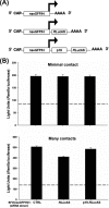
SFV-induced siRNA spread between U4.4 mosquito cells. (A) Replicons used to produce VLPs. eGFP was inserted between duplicated nsP2 cleavage sites in the nsP3/4 junction region of the nonstructural open reading frame. (B) RLuc expression plasmid pRL-CMV was electroporated into U4.4 cells (reporter cells), which were then mixed at a low density (minimal contact [top]) or high density (many contacts [bottom]) with U4.4 cells infected for 24 h with the VLPs SFV(nseGFP/H)1 (control replicon [CTRL]), SFV(nseGFP/H)1-RLucAS, or SFV(nseGFP/H)1-p19-RLucAS to allow siRNA accumulation (donor cells). RLuc activities were determined at 24 h postmixing. Dotted line, background. Each bar represents the mean of three replicates; error bars indicate standard deviations. Every experiment was repeated at least twice.
FIG. 7.
FIG. 7.
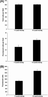
Scrape loading of siRNA and spread of fluorescein-labeled siRNA (Block-iT fluorescent oligonucleotide; Invitrogen). Scraping of cells from the surface using a cell scraper briefly results in damaged membranes through which RNA can be taken up (see Materials and Methods). U4.4 mosquito cells were scraped from the dish surface and immediately incubated with fluorescein-labeled siRNA for 5 h; scrape-loaded cells were then washed, counted, mixed with fresh U4.4 cells at a ratio of 1:10, and plated onto coverslips (A) or into wells (B). (A) Cells were fixed at 1 h (to allow cell attachment and recovery of normal morphology) and 6 h postplating; total cell numbers (top) and numbers of fluorescent cells (bottom) per field were counted (at magnifications of ×40 [10 fields] and ×20 [15 fields], respectively). (B) Alternatively, cells seeded into wells (in triplicate for each time point) were scraped and immediately analyzed by FACS; for each data point, 20,000 cells were gated, and fluorescent cells were counted. Numbers of fluorescent cells at 1 and 6 h postmixing are indicated. Bars represent the means, and error bars indicate standard deviations. Every experiment was repeated at least twice.
FIG. 8.
FIG. 8.
Spread of the RNAi signal between cells results in antiviral activity. Parallel cultures of U4.4 mosquito cells were noninfected (NIC) (A and B) or infected (MOI of 10) with VLPs containing the SFV replicons SFV(nseGFP/H)1 (control replicon [CTRL]) (A and B), SFV(nseGFP/H)1-NS1 (A), or SFV(nseGFP/H)1-p19 (B) and then incubated for 24 h to allow replicon-derived dsRNA/siRNA accumulation in donor cells. Replicons express eGFP from the nonstructural region (as described in the legend of Fig. 6) and no additional protein (control), influenza virus NS1 (dsRNA/siRNA-binding RNAi inhibitor in arthropod cells), or tombusvirus p19 (siRNA-binding protein) from the subgenomic promoter. After 24 h, replicon-containing or noninfected cells were mixed with U4.4 cells freshly infected (1 h p.i.) with RLuc-encoding SFV4(3H)-RLuc virus (A) or SFV(RLuc/H)1 VLPs (B) (MOI of 10) (reporter cells) at low (minimal contact) or high (many contacts) density. RLuc activity (indicating replication of virus) was measured 24 h postmixing. (C) Cell-to-cell spread of siRNA inhibits SFV4 production. U4.4 mosquito cells were noninfected (NIC) or infected (MOI of 10) with VLPs containing SFV replicons (as described above), SFV(nseGFP/H)1 (control replicon), or SFV(nseGFP/H)1-p19 and then incubated for 24 h to allow replicon-derived dsRNA/siRNA accumulation in donor cells. After 24 h, replicon-containing or noninfected donor cells were mixed with U4.4 cells freshly infected (1 h p.i.) with SFV4 (MOI of 1) at high density (many contacts). SFV4 production at 12 and 24 h postmixing is shown; virus titers (PFU/ml) were determined by plaque assay (as described in Materials and Methods). Each bar represents the mean of three replicates; error bars indicate standard deviations. Every experiment was repeated at least twice.
FIG. 9.
FIG. 9.
Replication of SFV encoding an RNAi inhibitor. (A) RLuc reporter SFV expressing tombusvirus siRNA-binding protein p19 from a duplicated subgenomic promoter [SFV4(3H)-RLuc-p19]. (B) Confluent U4.4 cells were infected at an MOI of 1 with SFV4(3H)-RLuc (Fig. 3) or SFV4(3H)-RLuc-p19 and transfected with RLuc or negative control (nc) siRNAs (concentration of 10 nM) for 5 h at 8 h p.i. RLuc activities were measured 24 h posttransfection. Results are expressed as a percentage of the control value (nontransfected, infected U4.4 cells [dotted line]). (C) Confluent U4.4 mosquito cells were infected at a high MOI (MOI of 10) or low MOI (MOI of 0.001) with SFV4(3H)-RLuc-p19 or SFV4(3H)-RLuc. Cells were lysed at 48 h p.i., and RLuc activities were determined. High MOI mimics initial infection as all cells are infected, while low MOI allows the virus to spread through the monolayer and undergo multiple rounds of infection. (D) Infection of U4.4 mosquito cells with SFV4(3H)-RLuc or SFV4(3H)-RLuc-p19 at high MOI (MOI of 10) or low MOI (MOI of 0.001). RNA was extracted at 48 h p.i., and virus genome copy numbers were determined by real-time qPCR (targeting a region of nsP3 as described in Materials and Methods). (E) U4.4 mosquito cells were infected with SFV4(3H)-RLuc or SFV4(3H)-RLuc-p19 at high MOI (MOI of 10) or low MOI (MOI of 0.001). Virus production (PFU/ml) was measured at 6, 24, and 48 h p.i. by plaque assay titration of supernatants (as described in Materials and Methods). •, SFV4(3H)-RLuc-p19; ○, SFV4(3H)-RLuc. Each bar represents the mean of three replicates (with the exception of real-time qPCR, where more replicates were analyzed) (see Materials and Methods); error bars indicate standard deviations. Every experiment was repeated at least twice.
FIG. 10.
FIG. 10.
Visualization of SFV spread. U4.4 mosquito cells were infected with SFV4(3H)-RLuc (A and B) or SFV4(3H)-RLuc-p19 (C and D) at a high MOI (MOI of 10) (A and C) or low MOI (MOI of 0.001) (B and D). Cells were fixed at 24 h p.i. and stained with an anti-nsP3 antibody (as described in Materials and Methods). Representative sections of cells are shown; selected replication complexes (bright focal staining) are indicated by arrows.

References

    1. Adelman, Z. N., I. Sanchez-Vargas, E. A. Travanty, J. O. Carlson, B. J. Beaty, C. D. Blair, and K. E. Olson. 2002. RNA silencing of dengue virus type 2 replication in transformed C6/36 mosquito cells transcribing an inverted-repeat RNA derived from the virus genome. J. Virol. 7612925-12933. - PMC - PubMed
    1. Aguilar, P. V., S. C. Weaver, and C. F. Basler. 2007. Capsid protein of eastern equine encephalitis virus inhibits host cell gene expression. J. Virol. 813866-3876. - PMC - PubMed
    1. Baulcombe, D. 2004. RNA silencing in plants. Nature 431356-363. - PubMed
    1. Billecocq, A., M. Vazeille-Falcoz, F. Rodhain, and M. Bouloy. 2000. Pathogen-specific resistance to Rift Valley fever virus infection is induced in mosquito cells by expression of the recombinant nucleoprotein but not NSs non-structural protein sequences. J. Gen. Virol. 812161-2166. - PubMed
    1. Blakqori, G., S. Delhaye, M. Habjan, C. D. Blair, I. Sanchez-Vargas, K. E. Olson, G. Attarzadeh-Yazdi, R. Fragkoudis, A. Kohl, U. Kalinke, S. Weiss, T. Michiels, P. Staeheli, and F. Weber. 2007. La Crosse bunyavirus nonstructural protein NSs serves to suppress the type I interferon system of mammalian hosts. J. Virol. 814991-4999. - PMC - PubMed

Publication types