. 2009 Jun;83(11):5375-87.
doi: 10.1128/JVI.00109-09. Epub 2009 Mar 18.

Evidence that productive human immunodeficiency virus type 1 assembly can occur in an intracellular compartment

Affiliations

Evidence that productive human immunodeficiency virus type 1 assembly can occur in an intracellular compartment

Anjali Joshi et al. J Virol. 2009 Jun.

Abstract

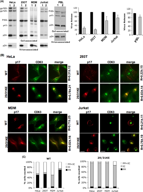
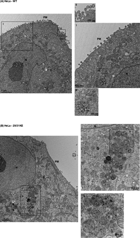
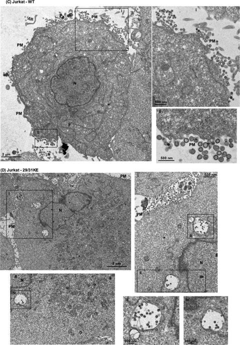
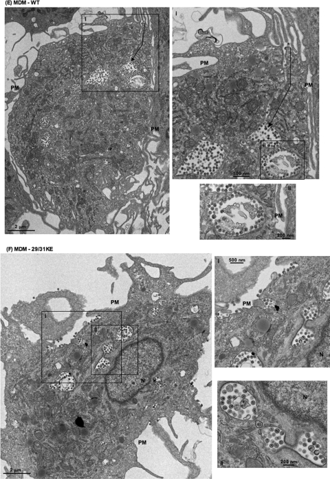
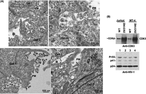
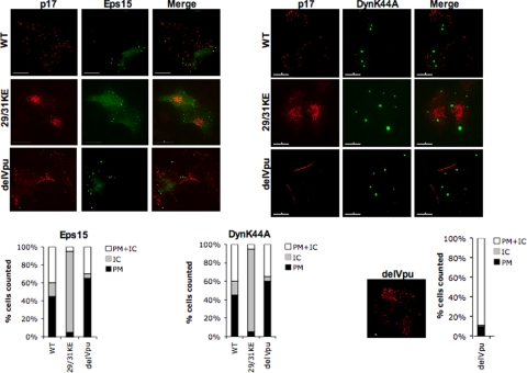
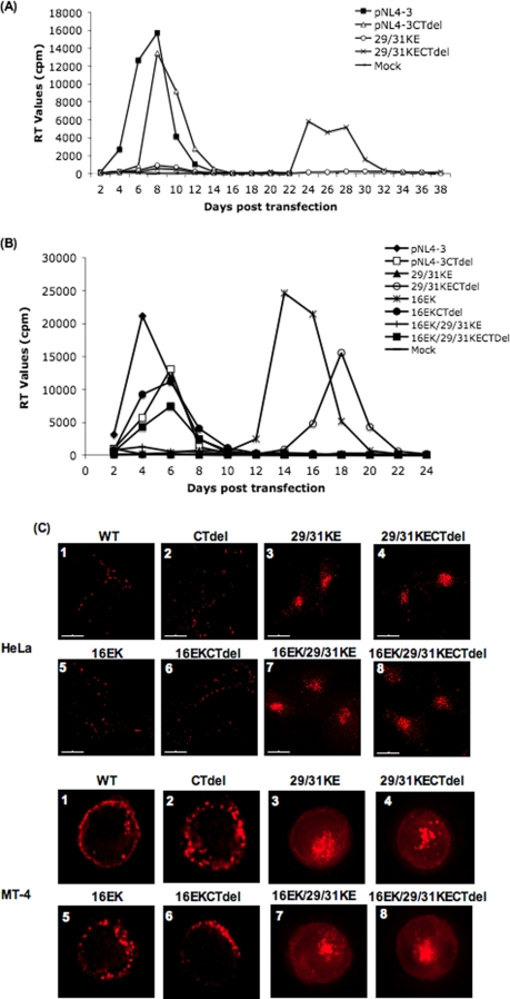
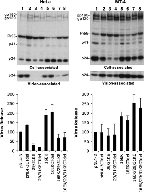
Human immunodeficiency virus type 1 (HIV-1) assembly occurs predominantly at the plasma membrane of infected cells. The targeting of assembly to intracellular compartments such as multivesicular bodies (MVBs) generally leads to a significant reduction in virus release efficiency, suggesting that MVBs are a nonproductive site for HIV-1 assembly. In the current study, we make use of an HIV-1 Gag-matrix mutant, 29/31KE, that is MVB targeted. We previously showed that this mutant is severely defective for virus particle production in HeLa cells but more modestly affected in primary macrophages. To more broadly examine the consequences of MVB targeting for virus production, we investigated 29/31KE particle production in a range of cell types. Surprisingly, this mutant supported highly efficient assembly and release in T cells despite its striking MVB Gag localization. Manipulation of cellular endocytic pathways revealed that unlike Vpu-defective HIV-1, which demonstrated intracellular Gag localization as a result of Gag endocytosis from the plasma membrane, 29/31KE mutant Gag was targeted directly to an MVB compartment. The 29/31KE mutant was unable to support multiple-round replication; however, this defect could be reversed by truncating the cytoplasmic tail of the transmembrane envelope glycoprotein gp41 and by the acquisition of a 16EK change in matrix. The 16EK/29/31KE matrix mutant replicated efficiently in the MT-4 T-cell line despite maintaining an MVB-targeting phenotype. These results indicate that MVB-targeted Gag can be efficiently released from T cells and primary macrophages, suggesting that under some circumstances, late endosomal compartments can serve as productive sites for HIV-1 assembly in these physiologically relevant cell types.


Figures

FIG. 1.
FIG. 1.
Release of the MVB-targeted MA mutant 29/31KE is severely impaired in HeLa and 293T cells but is efficient in MDM and Jurkat T cells. The indicated cell types were infected with RT-normalized stocks of VSV-G-pseudotyped WT or 29/31KE mutant virus. (A, left) Twenty-four hours postinfection, cells were metabolically radiolabeled with [35S]Met/Cys, and cell and virus lysates were immunoprecipitated with HIV-Ig and resolved by SDS-PAGE. (Right) Bands were quantified by fluorography, and virus release efficiency was calculated. Lanes 1 and black bars, WT; lanes 2 and gray bars, 29/31KE. Error bars represent means ± standard deviations (n = 3 for HeLa and 293T cells; n = 5 for MDMs). For analysis of virus release in PBLs, cell and virus lysates were subjected to SDS-PAGE and then immunoblotted with HIV-Ig. Bands were quantified by using the AlphaInnotech digital imager, and virus release efficiency was calculated. (B) Twenty-four hours postinfection, cells were fixed and labeled with anti-p17 (MA) and anti-CD63 Abs, followed by image acquisition using a DeltaVision RT deconvolution microscope. Colocalization between Gag and CD63 was determined using the SoftWorx colocalization module. R is the Pearson coefficient of correlation ± standard deviations averaged for 10 to 15 cells. (C) Quantification of Gag localization at the PM or intracellular (IC) sites for WT or 29/31KE Gag in an average of 10 to 15 cells. Gag localization was scored as PM or intracellular only if >90% of the staining was observed on the PM or in internal compartments.
FIG. 2.
FIG. 2.
The 29/31KE mutant assembles predominantly in an internal compartment in all cell types tested. Shown is EM analysis of WT HIV-1 and the 29/31KE mutant in HeLa (A and B), Jurkat (C and D), and MDM (E and F) cells. Cells were infected with VSV-G-pseudotyped WT HIV-1 or the 29/31KE mutant. Cells were fixed 24 to 48 h postinfection and analyzed by EM. N, nucleus. Insets represent magnified portions of the image with corresponding numbers (I, II, and III). In E, the arrow indicates the channel connecting the internal virus-containing compartment and the PM.
FIG. 2.
FIG. 2.
The 29/31KE mutant assembles predominantly in an internal compartment in all cell types tested. Shown is EM analysis of WT HIV-1 and the 29/31KE mutant in HeLa (A and B), Jurkat (C and D), and MDM (E and F) cells. Cells were infected with VSV-G-pseudotyped WT HIV-1 or the 29/31KE mutant. Cells were fixed 24 to 48 h postinfection and analyzed by EM. N, nucleus. Insets represent magnified portions of the image with corresponding numbers (I, II, and III). In E, the arrow indicates the channel connecting the internal virus-containing compartment and the PM.
FIG. 3.
FIG. 3.
Evidence that released 29/31KE virions assemble in an internal CD63+ compartment. (A) Jurkat T cells were infected with VSV-G-pseudotyped 29/31KE virus stocks. Cells were fixed 24 to 48 h postinfection and analyzed by EM. Adjacent cells are indicated as cell 1 and cell 2. (B) Jurkat or MT-4 cells were infected with RT-normalized VSV-G-pseudotyped WT or 29/31KE virus stocks. Culture supernatants were harvested 24 h postinfection, ultracentrifuged, and analyzed for CD63 incorporation into virions by Western blotting (WB) (top). The blot was stripped and reprobed with HIV-Ig (bottom).
FIG. 4.
FIG. 4.
29/31KE Gag is targeted directly to MVBs rather than being internalized from the PM. HeLa cells were transfected with HIV-WT, 29/31KE, and delVpu molecular clones along with vectors expressing dominant-negative Eps15 or GFP-tagged dynamin-K44A (DynK44A). Cells were fixed 24 h posttransfection, stained with anti-p17 Ab, and subjected to immunofluorescence analysis. Graphs under each panel represent PM, intracellular (IC), or mixed PM and IC Gag localization in an average of 10 to 15 cells. The staining pattern for delVpu in the absence of any inhibitors along with the PM or IC localization pattern averaged from 15 cells is depicted as a graph at the bottom right.
FIG. 5.
FIG. 5.
The 29/31KE mutant replicates in the MT-4 T-cell line after truncation of the gp41 cytoplasmic tail and acquisition of the 16EK compensatory mutation despite being MVB localized. (A and B) MT-4 T cells were transfected with WT pNL4-3, pNL4-3 bearing the CTdel-144-2 gp41 truncation (CTdel), and/or the 29/31KE, 16EK, or 16EK/29/31KE MA mutation. Cells were split every 2 days, and supernatants were reserved at each time point for RT assay. (C) HeLa (top) or MT-4 (bottom) cells were infected with the indicated VSV-G-pseudotyped virus stocks. Cells were fixed 24 to 48 h postinfection and stained with anti-p17 Ab.
FIG. 6.
FIG. 6.
The 16EK mutation significantly increases the level of production of cell-free virus. HeLa and MT-4 cells expressing the indicated pNL4-3 molecular clones, encoding WT or truncated (CTdel) Env and WT or mutant MA domains, were metabolically labeled with [35S]Met/Cys. Cell and virion lysates were immunoprecipitated and analyzed as described in the Fig. 1 legend.

References

    1. Adamson, C. S., and E. O. Freed. 2007. Human immunodeficiency virus type 1 assembly, release, and maturation. Adv. Pharmacol. 55347-387. - PubMed
    1. Batonick, M., M. Favre, M. Boge, P. Spearman, S. Honing, and M. Thali. 2005. Interaction of HIV-1 Gag with the clathrin-associated adaptor AP-2. Virology 342190-200. - PubMed
    1. Bieniasz, P. D. 2006. Late budding domains and host proteins in enveloped virus release. Virology 34455-63. - PubMed
    1. Bryant, M., and L. Ratner. 1990. Myristoylation-dependent replication and assembly of human immunodeficiency virus 1. Proc. Natl. Acad. Sci. USA 87523-527. - PMC - PubMed
    1. Camus, G., C. Segura-Morales, D. Molle, S. Lopez-Verges, C. Begon-Pescia, C. Cazevieille, P. Schu, E. Bertrand, C. Berlioz-Torrent, and E. Basyuk. 2007. The clathrin adaptor complex AP-1 binds HIV-1 and MLV Gag and facilitates their budding. Mol. Biol. Cell 183193-3203. - PMC - PubMed

Publication types

Substances

LinkOut - more resources