. 2009 May;20(9):2495-507.
doi: 10.1091/mbc.e08-09-0969. Epub 2009 Mar 18.

Down-regulation of the met receptor tyrosine kinase by presenilin-dependent regulated intramembrane proteolysis

Affiliations

Down-regulation of the met receptor tyrosine kinase by presenilin-dependent regulated intramembrane proteolysis

Bénédicte Foveau et al. Mol Biol Cell. 2009 May.

Abstract

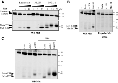
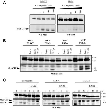
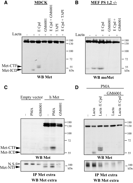
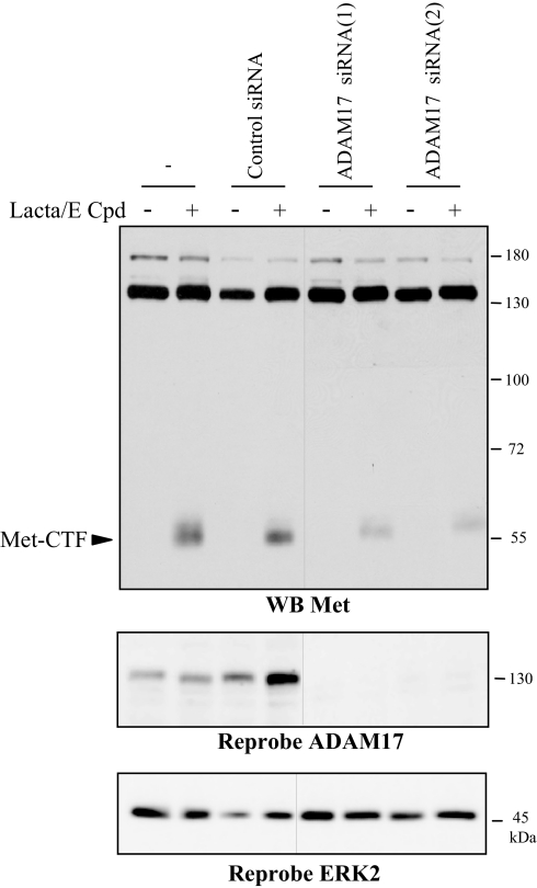
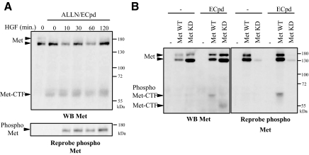
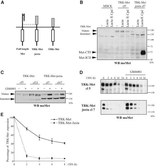
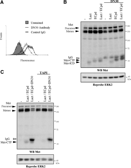
Hepatocyte growth factor/scatter factor (HGF/SF) acts through the membrane-anchored Met receptor tyrosine kinase to induce invasive growth. Deregulation of this signaling is associated with tumorigenesis and involves, in most cases, overexpression of the receptor. We demonstrate that Met is processed in epithelial cells by presenilin-dependent regulated intramembrane proteolysis (PS-RIP) independently of ligand stimulation. The proteolytic process involves sequential cleavage by metalloproteases and the gamma-secretase complex, leading to generation of labile fragments. In normal epithelial cells, although expression of cleavable Met by PS-RIP is down-regulated, uncleavable Met displayed membrane accumulation and induced ligand-independent motility and morphogenesis. Inversely, in transformed cells, the Met inhibitory antibody DN30 is able to promote Met PS-RIP, resulting in down-regulation of the receptor and inhibition of the Met-dependent invasive growth. This demonstrates the original involvement of a proteolytic process in degradation of the Met receptor implicated in negative regulation of invasive growth.


Figures

Figure 1.
Figure 1.
Stabilization of Met fragments by proteasome inhibitors. (A) MDCK epithelial cells were treated for 5 h with increasing concentrations of lactacystin or for 10 h with increasing concentrations of ALLN or MG132. (B) HeLa cells were treated for 5 h with lactacystin or for 10 h with ALLN or MG132. (C) HeLa cells were treated for 5 h with lactacystin or for 10 h with ALLN or MG132 and for 45 min with 100 ng/ml PMA. (A–C) For each condition, the same amount of protein was resolved by 10% SDS-PAGE and analyzed by Western blotting with antibodies against the kinase domain of Met (WB Met) or against the extracellular domain of Met (WB Met extra). The positions of prestained molecular weight markers are indicated. Arrows indicate positions of precursor and mature full-length Met, Met-CTF, and Met-ICD.
Figure 2.
Figure 2.
γ-Secretase inhibition prevents the Met-CTF-to-Met-ICD transition. (A) MDCK and HeLa cells were treated or not overnight with increasing concentrations of E compound. (B) Wild-type mouse embryonic fibroblasts (MEF PS WT), presenilin 1–deficient MEF (PS1−/−), presenilin 2–deficient MEF (PS2 −/−), and presenilin 1– and 2–deficient MEF (PS1,2 −/−) were treated or not overnight with 1 μM E compound (E-Cpd). The following day, the cells were treated or not for 5 h with 10 μM lactacystin. (C) MDCK epithelial cells were treated or not overnight with 1 μM E compound. The following day the cells were treated for 5 h with increasing concentrations of lactacystin or for 10 h with increasing concentrations of ALLN or MG132. (A–C) For each condition, the same amount of protein was resolved by 10% SDS-PAGE and analyzed by Western blotting with an antibody directed against the kinase domain of Met (WB Met) or the C-terminal domain of mouse Met for the Western blot produced with MEF extracts (WB moMet). Arrows indicate positions of Met-CTF and Met-ICD.
Figure 3.
Figure 3.
Metalloprotease inhibitors prevent generation of both Met-CTF and Met-ICD. (A) MDCK cells were left untreated (−) or were treated overnight with 1 μM E compound and/or 25 μM GM6001 and 50 μM TAPI-1. The following day the cells were treated for 5 h with 10 μM lactacystin. (B) MEF PS1,2 −/− were left untreated or treated overnight with 1 μM E compound and/or 25 μM GM6001. The following day the cells were treated for 5 h with 10 μM lactacystin. (C) MDCK cells transiently transfected with either the empty vector or a vector expressing human Met (h Met) were treated or not overnight with 25 μM GM6001. The following day, the cells were treated for 45 min with 100 ng/ml PMA. The culture medium was collected and immunoprecipitation was performed with an antibody directed against the extracellular domain of human Met. (D) HeLa cells were treated or not overnight with 1 μM E compound and/or 25 μM GM6001. The following day, the cells were treated for 5 h with 10 μM lactacystin and for 45 min with 100 ng/ml PMA. The culture medium was collected, and immunoprecipitation was performed with an antibody directed against the extracellular domain of human Met. (A–D) For each condition, the same amount of protein was resolved by 10% SDS-PAGE or 7% SDS-PAGE for IP and analyzed by Western blotting with different anti-Met antibodies as indicated. Arrows indicate positions of Met-CTF, Met-ICD, and Met-NTF.
Figure 4.
Figure 4.
ADAM-17 is involved in Met shedding. MCF10A cells were transfected or not with siRNA control or targeting the ADAM-17. Cells were then treated or not overnight with 1 μM E compound and the following day 5 h with 10 μM lactacystin. Proteins were resolved by 10% SDS-PAGE and analyzed by Western blotting with antibodies directed against the kinase domain of Met (WB Met). The filter was stripped and reprobed with an antibody directed against the ADAM-17 and against ERK2 to assess the loading. The two parts of the panels were on the same gel. Arrows indicate the position of Met-CTF.
Figure 5.
Figure 5.
HGF/SF does not influence PS-RIP of Met. (A) MDCK cells were treated or not overnight with 1 μM E compound and the following day 10 h with ALLN. Cells were then stimulated with HGF/SF (30 ng/ml) at the indicated time. (B) MDCK cells transiently transfected with either the empty vector or the vector expressing full-length human wild-type Met (Met WT) or kinase-dead Met (Met KD) were treated or not overnight with 1 μM E compound. (A and B) Proteins were resolved by 10% SDS-PAGE and analyzed by Western blotting with antibodies directed against the kinase domain of Met (WB Met). The filter was stripped and reprobed with an antibody directed against the phosphorylated tyrosine residues of the kinase domain of Met. Arrows indicate the positions of Met, Met-CTF and phosphorylated Met and Met-CTF.
Figure 6.
Figure 6.
PS-RIP regulates the stability of TRK-Met-juxta. (A) Schematic representation of full-length Met, TRK-Met (consisting of the extracellular portion of TRKA fused to the transmembrane and intracellular domains of Met), and TRK-Met-juxta, possessing 50 additional amino acids of the extracellular juxtamembrane domain of Met. (B) MDCK cells transfected with either the empty vector or the vector expressing TRK-Met (clones 5) or TRK-Met-juxta (clones 7) were treated or not overnight with 1 μM E compound. The following day the cells were treated for 5 h with lactacystin (10 μM) before lysis. (C) MDCK cells stably expressing TRK-Met or TRK-Met-juxta were treated or not overnight with 25 μM GM6001. The following day the cells were lysed. (D) MDCK cells stably expressing TRK-Met or TRK-Met-juxta were treated or not overnight with 25 μM GM6001. The following day the cells were treated with 25 μg/ml CHX for the indicated time. (B–D) Proteins were resolved by 10% SDS-PAGE and analyzed by Western blotting with antibodies directed against the C-terminal domain of mouse Met (WB moMet). Arrows indicate the positions of the mature and immature forms of TRK-Met and TRK-Met-juxta, Met-CTF, and Met-ICD. (E) MDCK cells expressing TRK-Met or TRK-Met-juxta were treated or not with 25 μg/ml CHX for the indicated time. Luminescence from the Western blot was captured with a CCD camera and the expression levels of mature TRK-Met were quantified. The percentage of expression was calculated using the untreated control as the reference (n = 3, ± SD).
Figure 7.
Figure 7.
PS-RIP occurs on the mature TRK-Met chimera at the membrane. (A) MDCK cells stably expressing TRK-Met-juxta or TRK-Met were treated or not overnight with 25 μM GM6001. Nuclei were detected by Hoechst staining (Hoechst, blue staining), and immunofluorescence staining was done with an anti-mouse Met antibody (Anti-mouse Met, red staining). Magnification, ×100. (B) TRK-Met– and TRK-Met-juxta–expressing cells were stained (red, open histogram) or not (black, filled histogram) with a phycoerythrin-coupled antibody directed against the extracellular domain of TRKA, and fluorescence was analyzed by flow cytometry. (C) TRK-Met– and TRK-Met-juxta–expressing cells were treated or not overnight with 2.5 μM SU11274. Immunoprecipitation was performed with an anti-mouse Met antibody. Proteins were resolved by 10% SDS-PAGE and analyzed by Western blotting with an antibody against phosphorylated tyrosine residues of the kinase domain of Met. The filter was stripped and reprobed with an antibody directed against mouse Met. Arrows indicate the positions of the mature and immature forms of TRK-Met and TRK-Met-juxta.
Figure 8.
Figure 8.
PS-RIP of TRK-Met chimera prevents its basal activation and the induction of invasive growth. (A) Scattering from cell islets. MDCK stably expressing TRK-Met and TRK-Met-juxta were seeded at low density. The next day the cells were cultured in the presence or absence of 2.5 μM SU11274 and/or 100 ng/ml NGF. Magnification, ×40 (B) MDCK stably expressing TRK-Met and TRK-Met-juxta were seeded at low density. The next day, the cells were fixed and stained for F-actin with phalloidin. Magnification, ×100. (C) Morphogenesis on Matrigel gels. MDCK stably expressing TRK-Met and TRK-Met-juxta were cultured on Matrigel gels. The following day, cells were incubated with or without SU11274 and/or NGF. Magnification, ×60. (D) MDCK stably expressing TRK-Met and TRK-Met-juxta, cultured in invasion chambers, were treated or not with 5 μM SU11274, a Met kinase inhibitor. The relative number of infiltrated cells was determined after staining of nuclei with Hoechst (n = 3, ± SD).
Figure 9.
Figure 9.
Induction of the Met PS-RIP by the Met inhibitory antibody DN30. (A) MDA-MB231 cells were treated 48 h with 40 μg/ml DN30 antibody (open histogram, thin line) or nonrelevant IgG (open histogram, thick line). The cells were then incubated or not (filled histogram) with an antibody directed against the extracellular domain of Met and fluorescein-conjugated antibody. The fluorescence was analyzed by flow cytometry. (B) MDA-MB231 cells were treated or not with 40 μg/ml DN30 antibody. The following day the cells were incubated or not overnight with 1 μM E compound and then treated for 5 h with 10 μM lactacystin. (C) MDA-MB231 cells were treated or not with 40 μg/ml DN30 antibody. The following day the cells were treated or not overnight with 1 μM E compound and 50 μM TAPI-1. The cells were then treated for 5 h with 10 μM lactacystin. (B and C) Proteins were resolved by 10% SDS-PAGE and analyzed by Western blotting with antibodies directed against the kinase domain of Met (WB Met). The filter was stripped and reprobed with an antibody directed against ERK2. Arrows indicate the positions of precursor and mature full-length Met, Met-CTF, Met ICD, and IgG from DN30 antibody.

References

    1. Birchmeier C., Birchmeier W., Gherardi E., Vande Woude G. F. Met, metastasis, motility and more. Nat. Rev. Mol. Cell Biol. 2003;4:915–925. - PubMed
    1. Bladt F., Riethmacher D., Isenmann S., Aguzzi A., Birchmeier C. Essential role for the c-met receptor in the migration of myogenic precursor cells into the limb bud. Nature. 1995;376:768–771. - PubMed
    1. Borowiak M., Garratt A. N., Wustefeld T., Strehle M., Trautwein C., Birchmeier C. Met provides essential signals for liver regeneration. Proc. Natl. Acad. Sci. USA. 2004;101:10608–10613. - PMC - PubMed
    1. Brou C., Logeat F., Gupta N., Bessia C., LeBail O., Doedens J. R., Cumano A., Roux P., Black R. A., Israel A. A novel proteolytic cleavage involved in Notch signaling: the role of the disintegrin-metalloprotease TACE. Mol. Cell. 2000;5:207–216. - PubMed
    1. Comoglio P. M., Giordano S., Trusolino L. Drug development of MET inhibitors: targeting oncogene addiction and expedience. Nat. Rev. Drug Discov. 2008;7:504–516. - PubMed

Publication types

MeSH terms

LinkOut - more resources