. 2009 May;150(1):402-15.
doi: 10.1104/pp.109.135624. Epub 2009 Mar 18.

Grapevine MATE-type proteins act as vacuolar H+-dependent acylated anthocyanin transporters

Affiliations

Grapevine MATE-type proteins act as vacuolar H+-dependent acylated anthocyanin transporters

Camila Gomez et al. Plant Physiol. 2009 May.

Abstract

In grapevine (Vitis vinifera), anthocyanins are responsible for most of the red, blue, and purple pigmentation found in the skin of berries. In cells, anthocyanins are synthesized in the cytoplasm and accumulated into the vacuole. However, little is known about the transport of these compounds through the tonoplast. Recently, the sequencing of the grapevine genome allowed us to identify genes encoding proteins with high sequence similarity to the Multidrug And Toxic Extrusion (MATE) family. Among them, we selected two genes as anthocyanin transporter candidates and named them anthoMATE1 (AM1) and AM3. The expression of both genes was mainly fruit specific and concomitant with the accumulation of anthocyanin pigment. Subcellular localization assays in grapevine hairy roots stably transformed with AM1 or AM3green fluorescent protein fusion protein revealed that AM1 and AM3 are primarily localized to the tonoplast. Yeast vesicles expressing anthoMATEs transported acylated anthocyanins in the presence of MgATP. Inhibitor studies demonstrated that AM1 and AM3 proteins act in vitro as vacuolar H(+)-dependent acylated anthocyanin transporters. By contrast, under our experimental conditions, anthoMATEs could not transport malvidin 3-O-glucoside or cyanidin 3-O-glucoside, suggesting that the acyl conjugation was essential for the uptake. Taken together, these results provide evidence that in vitro the two grapevine AM1 and AM3 proteins mediate specifically acylated anthocyanin transport.


Figures

Figure 1.
Figure 1.
Phylogenetic tree of the predicted amino acid sequences of grapevine MATE proteins, Arabidopsis TT12 (Q9LYT3), and tomato MTP77 (AAQ55183). The asterisks indicate genes selected as being putative anthocyanin transporters. In the accession numbers of grapevine sequences annotated in the Genoscope grape genome browser (http://www.genoscope.cns.fr/vitis/; data obtained from the 8-fold coverage of the genome), “GSVIVP000” was simplified to “VvM.” Relative similarities between sequences (proportional to horizontal branch lengths) are drawn to scale.
Figure 2.
Figure 2.
Genomic structures of anthoMATE genes in scaffold_91 of the Genoscope genome browser database (http://www.genoscope.cns.fr). The black boxes indicate exons. In the accession numbers annotated in the Genoscope grape genome browser (http://www.genoscope.cns.fr/vitis/; data obtained from the 8-fold coverage of the genome), “GSVIVT000” was simplified to “VvM.”
Figure 3.
Figure 3.
Alignment of AM1 and AM3 proteins with Arabidopsis TT12 and tomato MTP77 using the ClustalW program with default parameters. The MatE domain (PF01554) using the Pfam database is shown with gray background. The putative transmembrane segments (TM1–TM12) of anthoMATE proteins, using the HMMTOP program, are delimited by thick lines above the sequences. Conserved residues in all sequences are shown with black background. Conserved protein domains (D1–D5) are represented by thin lines below the sequences. The anthoMATE motif used in antibody synthesis is indicated by the dotted line.
Figure 4.
Figure 4.
Subcellular localization of anthoMATE proteins after stable expression in grapevine hairy roots. Confocal microscopy images show grapevine root epidermal cells expressing nontargeted GFP protein (A), AM1∷GFP fusion (B and D), and AM3∷GFP fusion (C and E). Cells were plasmolyzed with 0.3 m sorbitol, and DNA was stained blue with DAPI and green with GFP. A, GFP fluorescence is visible in the cytoplasm. B to E, GFP fluorescence is visible on the tonoplast and in membrane structures attached to the nucleus (indicated by asterisks). N, Nucleus; V, vacuole. D and E show magnified images corresponding to the boxed regions in B and C, respectively. The image in C overlaid by differential interference contrast transmitted light is shown in Supplemental Figure S1B. Bars = 5 μm in A to C and 2 μm in D and E.
Figure 5.
Figure 5.
Quantitative real-time PCR expression profiling of AM1 and AM3. Transcript levels of AM1 (A) and AM3 (B) during berry development (Syrah) and transcript levels of AM1 (C) and AM3 (D) in various grapevine organs and different tissues of mature berries are shown. The arrows indicate the véraison. Gene expression was normalized with VvEF1α. All data are means of three replicates with error bars indicating sd.
Figure 6.
Figure 6.
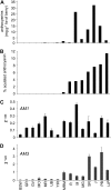
A, Anthocyanin content of mature berries from different cultivars. fw, Fresh weight. B, Acylated anthocyanin (expressed as percentage of total anthocyanin) in mature berries from different cultivars. C and D, Quantitative real-time PCR expression profiling of AM1 (C) and AM3 (D) in mature berries obtained from different cultivars. Gene expression was normalized with VvEF1α. All data are means of three replicates with error bars indicating sd. Cultivar names are as follows: BSW, Buckland Sweet Water; GR, Grec Rouge; GV, Goher Valtozo; MO, Muscat Ottonel; MR, Moscatel Rosado; UB, Ugni Blanc; YIR, Yai Izioum Rosovy; MRM, Muscat Rouge de Madeire; R, Rousaïtis; M, Molinara; MG, Molinera Gorda; SR, Sensit Rouge; J, Joubertin; PB, Petit Boushet; and LlP, Lledoner Pelut.
Figure 7.
Figure 7.
AM1- and AM3-mediated transport of acylated anthocyanins in yeast microsomal vesicles. A and B, Absorption scans of different substrates recovered from filters after a 1-min transport experiment in the presence of MgATP with AM1-containing vesicles (A) and AM3-contaning vesicles (B). Substrates used were as follows: acylated anthocyanin mixture (AAM; solid lines); C3G (dotted gray lines); and M3G (dotted black lines). AU, Absorbance units. C and D, Absorption scans of acylated anthocyanins washed off filters after the transport experiment with AM1-containing vesicles (C) and AM3-containing vesicles (D), representing the vesicle-associated amount of acylated anthocyanin. Transport was stopped by filtration after 30 s (dotted lines) or 60 s (solid lines). NEV, Transport experiments performed with vesicles isolated from empty vector control-transformed yeast. All transport experiments were performed in the presence of MgATP, except transport experiments after 1 min with AM-containing vesicles, which were performed with and without ATP (w/o ATP), indicated by solid gray lines. E, HPLC analysis of the acylated anthocyanins recovered from filters after a 1-min transport experiment with AM1-containing vesicles and AM3-contaning vesicles with ATP (ATP+) or without ATP (w/o ATP). Peak 1, Delphinidin 3-p-coumaroylglucosyl; peak 2, malvidin 3-acetylglucosyl; peak 3, cyanidin 3-p-coumaroylglucosyl; peak 4, petunidin 3-p-coumaroylglucosyl; peak 5, peonidin 3-p-coumaroylglucosyl; and peak 6, malvidin 3-p-coumaroylglucosyl.
Figure 8.
Figure 8.
Quantification of MgATP-dependent uptake activity of acylated anthocyanins by AM1 and AM3. Bars show mean values of the difference between the experiments with or without the addition of MgATP ± confidence interval at 0.95. Values shown are averages of eight transport experiments performed with independent vesicle preparations. Bars associated with the same letter indicate that there are no significant differences (P > 0.05) by Student's t test.

References

    1. Ageorges A, Fernandez L, Vialet S, Merdinoglu D, Terrier N, Romieu C (2006) Four specific isogenes of the anthocyanin metabolic pathway are systematically co-expressed with the red colour of grape berries. Plant Sci 170 372–383
    1. Alfenito MR, Souer E, Goodman CD, Buell R, Mol J, Koes R, Walbot V (1998) Functional complementation of anthocyanin sequestration in the vacuole by widely divergent glutathione S-transferases. Plant Cell 10 1135–1149 - PMC - PubMed
    1. Archetti M (2000) The origin of autumn colours by coevolution. J Theor Biol 205 625–630 - PubMed
    1. Blanc G, Wolfe KH (2004) Functional divergence of duplicated genes formed by polyploidy during Arabidopsis evolution. Plant Cell 16 1679–1691 - PMC - PubMed
    1. Boss PK, Davies C, Robinson SP (1996) Analysis of the expression of anthocyanin pathway genes in developing Vitis vinifera L. cv Shiraz grape berries and the implications for pathway regulation. Plant Physiol 111 1059–1066 - PMC - PubMed

Publication types

MeSH terms