. 2009 Apr 23;113(16):4045-59.
doi: 10.1021/jp810375d.

A tale of tails: how histone tails mediate chromatin compaction in different salt and linker histone environments

Affiliations

A tale of tails: how histone tails mediate chromatin compaction in different salt and linker histone environments

Gaurav Arya et al. J Phys Chem A. .

Abstract

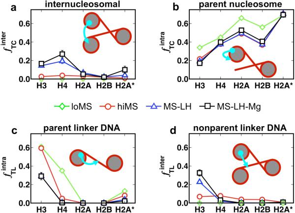
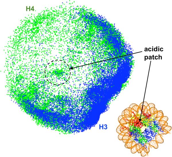
To elucidate the role of the histone tails in chromatin compaction and in higher-order folding of chromatin under physiological conditions, we extend a mesoscale model of chromatin (Arya, Zhang, and Schlick. Biophys. J. 2006, 91, 133; Arya and Schlick. Proc. Natl. Acad. Sci. U.S.A. 2006, 103, 16236) to account for divalent cations (Mg(2+)) and linker histones. Configurations of 24-nucleosome oligonucleosomes in different salt environments and in the presence and absence of linker histones are sampled by a mixture of local and global Monte Carlo methods. Analyses of the resulting ensembles reveal a dynamic synergism between the histone tails, linker histones, and ions in forming compact higher-order structures of chromatin. In the presence of monovalent salt alone, oligonucleosomes remain relatively unfolded, and the histone tails do not mediate many internucleosomal interactions. Upon the addition of linker histones and divalent cations, the oligonucleosomes undergo a significant compaction triggered by a dramatic increase in the internucleosomal interactions mediated by the histone tails, formation of a rigid linker DNA "stem" around the linker histones' C-terminal domains, and reduction in the electrostatic repulsion between linker DNAs via sharp bending in some linker DNAs caused by the divalent cations. Among all histone tails, the H4 tails mediate the most internucleosomal interactions, consistent with experimental observations, followed by the H3, H2A, and H2B tails in decreasing order. Apart from mediating internucleosomal interactions, the H3 tails also contribute to chromatin compaction by attaching to the entering and exiting linker DNA to screen electrotatic repulsion among the linker DNAs. This tendency of the H3 tails to attach to linker DNA, however, decreases significantly upon the addition of linker histones due to competition effects. The H2A and H2B tails do not mediate significant internucleosomal interactions but are important for mediating fiber/fiber intractions, especially in relatively unfolded chromatin in monovalent salt environments.


Figures

Figure 1
Figure 1
Mesoscale modeling of linker-histone bound oligonucleosomes. The nucleosome core is modeled as an irregular-shaped rigid body with 300 pseudo charges on it surface. The linker DNA is treated using the discrete version of the worm-like chain model. The linker histone is treated as charged beads connected rigidly to the nucleosome and the histone tails are treated using the protein bead model. The bottom figure at center shows the final integrated mesoscale model.
Figure 2
Figure 2
Mesoscale oligonucleosome model showing the: (a) assembly of oligonucleosome motifs into a chain; (b) the entering and exiting linker DNA geometry at the nucleosome core; and (c) the linker-DNA/nucleosome mechanics in terms of individual coordinate systems for the linker DNA beads and the nucleosome core.
Figure 3
Figure 3
Sedimentation coefficients of 12-unit oligonucleosomes with regular tails and neutralized tails at the four conditions: loMS, hiMS, MS-LH, and MS-LH-Mg (see Table 2). The open circles represent experimental results.,,
Figure 4
Figure 4
Representative oligonucleosomes obtained from our simulation ensemble highlighting the differences in the global morphology and internal structure of chromatin at the four conditions investigated in this study: (a) low monovalent salt (loMS); (b) high monovalent salt (hiMS); (c) high monovalent salt + linker histones (MS-LH); and (d) high monovalent salt + linker histone + magnesium cations (MS-LH-Mg). For clarity, a 12-unit oligonucleosome is presented in (a) and 24-unit oligonucleosomes are presented in (b-d). In (c,d), odd and even numbered nucleosome cores are colored white and blue, respectively, to highlight the predominant i ± 2 interactions; and severely bent linker DNAs are colored green, as characterized by an angle of bending greater than 90°. This bending angle is defined by the angle formed between the linker DNA exiting one nucleosome and entering the next nucleosome.
Figure 5
Figure 5
Positional distribution of histone tails under different conditions: low monovalent salt (a,d); high monovalent salt (b,e); and high monovalent salt with bound linker histones (c,f). The upper panel (a-c) represents the histone tail positions projected onto the nucleosomal plane of the parent nucleosomes, and the lower panel (e-f) represents the positions projected onto the dyad plane of the parent nucleosomes. The tails are colored as: H2A, yellow; H2B, red; H3, blue; and H4, green. The nucleosome boundaries are indicated by the solid black lines.
Figure 6
Figure 6
Histone tail interactions within chromatin captures in terms of the frequency with which they mediate: (a) internucleosomal interactions; (b) attach to parent nucleosomes; (c) attach to linker DNA associated with the parent nucleosome; and (d) linker DNA not associated with the parent nucleosome. The results are presented for the four conditions: chromatin at low monovalent salt (green down triangles); high monovalent salt (red circles); high monovalent salt with linker histones (blue up triangles); and high monovalent salt with linker histone and Mg2+ (black squares), respectively. The frequencies are calculated as the number of times a histone tail attaches to the chromatin component divided by the total number of sampled tail configurations.
Figure 7
Figure 7
Distribution of the histone tails' (H3, blue and H4, green) attachment sites on the surface of non-parental nucleosomes for MS-LH-Mg conditions. The distributions have been projected onto the plane of the nucleosome. The black circle denotes the acidic patch. The figure in the lower right corner shows the atomistic nucleosome with the same color coding as Fig. 1.

References

    1. Felsenfeld G, Groudine M. Nature. 2003;421:448–453. - PubMed
    1. Horn PJ, Peterson CL. Science. 2002;297:1824–1827. - PubMed
    1. Tse C, Hansen JC. Biochemistry. 1997;36:11381–11388. - PubMed
    1. Hansen JC, Tse C, Wolffe AP. Biochemistry. 1998;37:17637–17641. - PubMed
    1. Gordon F, Luger K, Hansen JC. J. Biol. Chem. 2005;280:33701–3706. - PubMed

Publication types