Review
. 2009 Apr;156(8):1185-95.
doi: 10.1111/j.1476-5381.2009.00111.x. Epub 2009 Mar 9.

Neural KCNQ (Kv7) channels

Affiliations
Review

Neural KCNQ (Kv7) channels

David A Brown et al. Br J Pharmacol. 2009 Apr.

Abstract

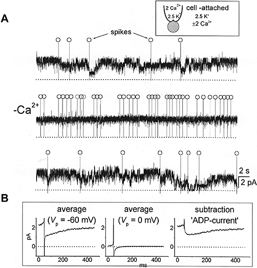
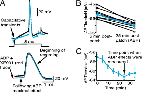
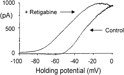
KCNQ genes encode five Kv7 K(+) channel subunits (Kv7.1-Kv7.5). Four of these (Kv7.2-Kv7.5) are expressed in the nervous system. Kv7.2 and Kv7.3 are the principal molecular components of the slow voltage-gated M-channel, which widely regulates neuronal excitability, although other subunits may contribute to M-like currents in some locations. M-channels are closed by receptors coupled to Gq such as M1 and M3 muscarinic receptors; this increases neuronal excitability and underlies some forms of cholinergic excitation. Muscarinic closure results from activation of phospholipase C and consequent hydrolysis and depletion of membrane phosphatidylinositol-4,5-bisphosphate, which is required for channel opening. Some effects of M-channel closure, determined from transmitter action, selective blocking drugs (linopirdine and XE991) and KCNQ2 gene disruption or manipulation, are as follows: (i) in sympathetic neurons: facilitation of repetitive discharges and conversion from phasic to tonic firing; (ii) in sensory nociceptive systems: facilitation of A-delta peripheral sensory fibre responses to noxious heat; and (iii) in hippocampal pyramidal neurons: facilitation of repetitive discharges, enhanced after-depolarization and burst-firing, and induction of spontaneous firing through a reduction of action potential threshold at the axon initial segment. Several drugs including flupirtine and retigabine enhance neural Kv7/M-channel activity, principally through a hyperpolarizing shift in their voltage gating. In consequence they reduce neural excitability and can inhibit nociceptive stimulation and transmission. Flupirtine is in use as a central analgesic; retigabine is under clinical trial as a broad-spectrum anticonvulsant and is an effective analgesic in animal models of chronic inflammatory and neuropathic pain.


Figures

Figure 1
Figure 1
Kv7/M-current in a rat sympathetic neuron. Records in (a) show M-currents recorded from a dissociated rat superior cervical sympathetic neuron with a perforated patch electrode on depolarizing the cell from −60 mV to −30 mV in 10 mV steps. Records in (b) show corresponding steady-state M-channel activity recorded through a cell-attached patch electrode on depolarizing the patch to the equivalent potentials to those in (a). (Adapted from figure 1 in Selyanko et al., 1992).
Figure 2
Figure 2
Inhibition of heteromeric Kv7.2/7.3 channels by the muscarinic agonist oxotremorine-M (Oxo-M). The channels were expressed in Chinese Hamster Ovary cells by KCNQ2 and KCNQ3 cDNA transfection. (A) Inhibition of whole-cell current recorded with a perforated patch electrode. Currents were generated by depolarizing the cell to −20 mV; records show deactivation of this steady outward current by 1 s hyperpolarizing steps to −50 mV. (B) Equivalent multi-channel activity recorded within a cell-attached membrane patch is inhibited by applying Oxo-M to the solution bathing the cell outside the patch, thereby activating muscarinic receptors remote from the Kv7.2/7.3 channels (‘remote inhibition’). (From Selyanko et al., 2000).
Figure 3
Figure 3
Effects of enhancing Kv/M-channel activity with retigabine and blocking Kv7/M-channels with XE991 on (left) membrane currents and (right) action potential generation in a dissociated rat sympathetic neuron. Records on the left show membrane currents recorded by depolarizing the cell to −20 mV (to activate Kv7/M-channels) then hyperpolarizing it to −50 mV (to deactivate the Kv7/M-current). Records in the presence of the drug are in red. Retigabine increases the outward Kv7/M-current at −20 mV and slows deactivation during hyperpolarization but accelerates reactivation. XE991 reduces the outward current and suppresses the slow deactivation relaxation, showing that Kv7/M-channels are blocked. Records on the right show responses to 4 s depolarizing currents up to 200 pA in 20 pA steps. Under normal conditions current injections ≥40 pA generated one (or sometimes two) action potentials at the beginning of the step. Retigabine suppressed spike generation whereas XE991 promoted repetitive spike discharges. Note: the starting membrane potential was held at −55 mV by constant current injection, offsetting the hyperpolarization and depolarization induced by retigabine and XE991 respectively (G.M. Passmore, unpublished).
Figure 4
Figure 4
Enhanced sensitivity of Aδ fibres to thermal stimulation by local application of XE991 in the rat isolated skin-nerve preparation. The preparation and recordings were obtained as described by Reeh (1986). Drugs were applied to the receptive field via the corium. Heat ramps from 30 to 46–47°C were applied for 90 s at 5 min intervals as shown. Single unit responses were recorded from a central portion of the saphenous nerve. Conduction velocity was 6.2 m·s−1. Records show second (control), fifth (+XE991 3 µmol·L−1) and eighth (wash) responses. Discharges per stimulus were 10, 52 and 10 respectively. (G.M. Passmore, unpublished).
Figure 5
Figure 5
Blocking Kv7/M-channels or suppressing Kv7.2 channel expression facilitates repetitive spike discharges in mouse CA1 pyramidal neurons. Records show spike discharges in response to 500 ms depolarizing currents, up to 200 pA, in hippocampal slices from (A–C) normal mice and (D–F) transgenic mice expressing a dominant-negative (pore-defective) Kv7.2 mutant. Neurons from normal mice (A) show strong spike-frequency adaptation; XE991 (B) greatly reduces adaptation, facilitating repetitive discharge. Neurons from the transgenic mice (D) show much less spike adaptation and already fire repetitively; this is not significantly enhanced by XE991 (E). (Adapted from Peters et al., 2005, with permission of Nature Publishing Group).
Figure 7
Figure 7
Transient Ca2+-dependent inhibition of Kv7/M-channels in dissociated hippocampal pyramidal neurons following spontaneous action potentials. (A) Cell-attached pipette recordings of multi-M channel activity obtained in 2 (upper and lower traces) and 0 (middle trace) mmole.L−1[Ca2+]. Spontaneous action potentials (‘spikes’) are marked (○). Note that action potentials are associated with intermittent channel closures in Ca2+-containing solution but not in Ca2+-free solution. (B) Ensembles of post-spike channel activity in Ca2+-containing solution recorded at −60 mV patch-potential (Vp), i.e., near-zero membrane potential (94 individual records) and 0 mV Vp, i.e., rest potential where most M-channels are shut (68 records). The latter was then subtracted from the former to yield (on the right) the net after-depolarizing current (ADP-current) resulting from spike-induced M-channel inhibition. (A.A.Selyanko, unpublished; data from Figures 11 and 12 in Selyanko & Sim, 1998, with permission of the Journal of Physiology.)
Figure 6
Figure 6
Blocking Kv7/M-channels enhances a spike after-depolarization and induces burst-firing in rat CA1 hippocampal pyramidal neurons. Normally a brief current injection induces a single action potential (Aa1,b1). In a2–a4, XE991 gradually depolarized the cell and induced burst-firing. This resulted from an enhanced after-depolarization (B). Row Ab1–b4 and panel C show that XE991 had the same effects even when the depolarization was prevented. (Adapted from Yue and Yaari, 2004, with permission of the Journal of Neuroscience).
Figure 8
Figure 8
Kv7/M-channel inhibition or disruption of Kv7/M-channel binding to ankyrin at the axon initial segment induces spontaneous action potential firing in hippocampal CA1 pyramidal neurons. Note: synaptic transmission was blocked by using a mixture of ionotropic and metabotropic glutamate and GABA antagonists. (A) Bath-applied XE991 (3 µmol·L−1) (a) depolarized the neuron, increased input resistance and facilitated repetitive firing in response to depolarizing current injections (a) and (b) induced spontaneous firing. (B) These effects were partly replicated by intracellular application of a peptide (ABP, ankyrin-binding peptide), designed to disrupt Kv7 channel binding to ankyrin at the axon initial segment, except that the peptide did not increase somatic input resistance (indicating that it did not affect somatic Kv7/M-channels). (Adapted from Shah et al., 2008).
Figure 9
Figure 9
Kv7/M-channels in the axon initial segment control action potential (AP) threshold in hippocampal CA1 pyramidal neurons: inclusion of ankyrin-binding peptide (ABP; see Figure 8) in the patch-pipette progressively reduces AP threshold. (A) Superimposed examples of single APs 5 (black trace) and 25 min (blue trace) post patching with ABP in the patch-pipette solution. Each AP was generated by a 5 ms depolarizing current injection from −70 mV, as shown in the schematic. The magnitude of the current injection was adjusted to ensure that the step produced only a single AP. The APs are also shown on an enhanced time scale below. Note that subsequent bath addition of XE991 (red trace) did not further reduce the AP threshold. (B) Average (blue squares) AP thresholds at the beginning and 25 min post patching with ABP in the internal pipette solution. Open black squares represent the effects in individual neurons. (C) Time course of the reduction in AP threshold with ABP. (From Shah et al., 2008).
Figure 10
Figure 10
Retigabine (10 µmol·L−1) produces a 20 mV shift of the Kv7/M-current in a sympathetic neuron. Ordinates: current, abscissae: membrane voltage. Currents were evoked by using −100–0 mV ramped voltage commands and Kv7/M-current measured as that current blocked by 10 µmol·L−1 linopirdine. As a result of the voltage shift, an outward current is generated at the resting potential (around −60 mV), leading to an average hyperpolarization of 9 mV. (From Tatulian et al., 2001).

References

    1. Adams PR, Jones SW, Pennefather P, Brown DA, Kock C, Lancaster B. Slow synaptic transmission in frog sympathetic ganglia. J Exp Biol. 1986;124:259–285. - PubMed
    1. Aiken SP, Lampe BJ, Murphy PA, Brown BS. Reduction of spike frequency adaptation and blockade of M-current in rat CA1 pyramidal neurones by linopirdine (DuP 996), a neurotransmitter release enhancer. Br J Pharmacol. 1995;115:1163–1168. - PMC - PubMed
    1. Aiken SP, Zaczek R, Brown BS. Pharmacology of the neurotransmitter release enhancer linopirdine (DuP 996), and insights into its mechanism of action. Adv Pharmacol. 1996;35:349–384. - PubMed
    1. Bentzen BH, Schmitt N, Calloe K, Dalby Brown W, Grunnet M, Olesen SP. The acrylamide (S)-1 differentially affects Kv7 (KCNQ) potassium channels. Neuropharmacology. 2006;51:1068–1077. - PubMed
    1. Blackburn-Munro G, Jensen BS. The anticonvulsant retigabine attenuates nociceptive behaviours in rat models of persistent and neuropathic pain. Eur J Pharmacol. 2003;460:109–116. - PubMed

Publication types

MeSH terms