Comparative Study
. 2009 May;33(5):791-8.
doi: 10.1111/j.1530-0277.2009.00897.x. Epub 2009 Mar 6.

Ethanol blocks adenosine uptake via inhibiting the nucleoside transport system in bronchial epithelial cells

Affiliations
Comparative Study

Ethanol blocks adenosine uptake via inhibiting the nucleoside transport system in bronchial epithelial cells

Diane S Allen-Gipson et al. Alcohol Clin Exp Res. 2009 May.

Abstract

Background: Adenosine uptake into cells by nucleoside transporters plays a significant role in governing extracellular adenosine concentration. Extracellular adenosine is an important signaling molecule that modulates many cellular functions via 4 G-protein-coupled receptor subtypes (A(1), A(2A), A(2B), and A(3)). Previously, we demonstrated that adenosine is critical in maintaining airway homeostasis and airway repair and that airway host defenses are impaired by alcohol. Taken together, we hypothesized that ethanol impairs adenosine uptake via the nucleoside transport system.

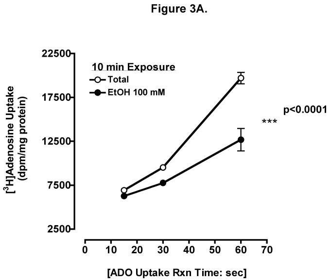
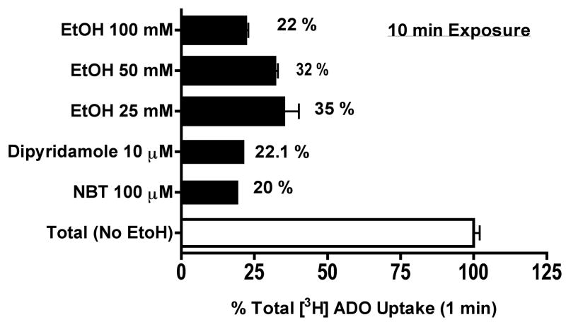
Methods: To examine ethanol-induced alteration on adenosine transport, we used a human bronchial epithelial cell line (BEAS-2B). Cells were preincubated for 10 minutes in the presence and absence of varying concentrations of ethanol (EtOH). In addition, some cells were pretreated with S-(4-Nitrobenzyl)-6-thioinosine (100 microM: NBT), a potent adenosine uptake inhibitor. Uptake was then determined by addition of [(3)H]-adenosine at various time intervals.

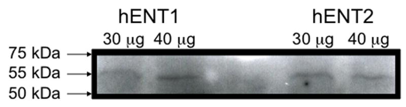
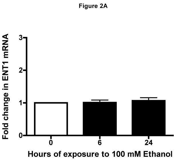
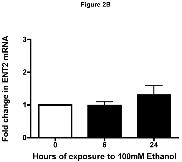
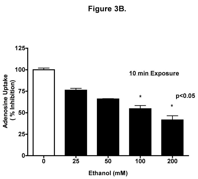
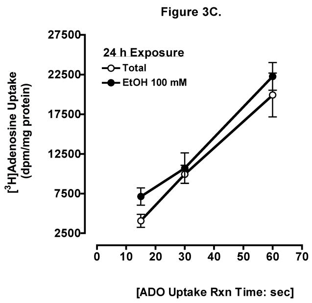
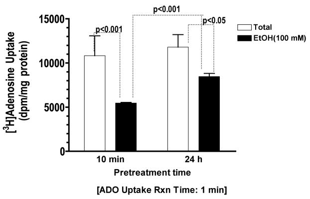
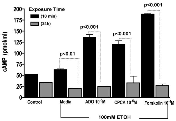
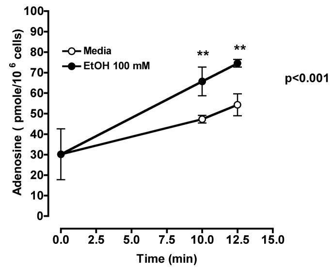
Results: Increasing EtOH concentrations resulted in increasing inhibition of adenosine uptake when measured at 1 minute. Cells pretreated with NBT effectively blocked adenosine uptake. In addition, short-term EtOH revealed increased extracellular adenosine concentration. Conversely, adenosine transport became desensitized in cells exposed to EtOH (100 mM) for 24 hours. To determine the mechanism of EtOH-induced desensitization of adenosine transport, cAMP activity was assessed in response to EtOH. Short-term EtOH exposure (10 minutes) had little or no effect on adenosine-mediated cAMP activation, whereas long-term EtOH exposure (24 hours) blocked adenosine-mediated cAMP activation. Western blot analysis of lysates from unstimulated BEAS-2B cells detected a single 55 kDa band indicating the presence of hENT1 and hENT2, respectively. Real-time RT-PCR of RNA from BEAS-2B revealed transcriptional expression of ENT1 and ENT2.

Conclusions: Collectively, these data reveal that acute exposure of cells to EtOH inhibits adenosine uptake via a nucleoside transporter, and chronic exposure of cells to EtOH desensitizes the adenosine transporter to these inhibitory effects of ethanol. Furthermore, our data suggest that inhibition of adenosine uptake by EtOH leads to an increased extracellular adenosine accumulation, influencing the effect of adenosine at the epithelial cell surface, which may alter airway homeostasis.


Figures

Figure 1
Figure 1. Western blot analysis of hENT1 and hENT2 expression
Representative Western blots identifying a single band at 55 kda of hENT1 and hENT2 proteins in crude extracts from human bronchial epithelial cells, BEAS-2B.
Figure 2
Figure 2. Identification of human equilibrative nucleoside transporters in human epithelial cells
Transcriptional Level of (A) hENT1 and of hENT2 (B) were quantified using real-time PCR in RNA isolated from BEAS-2B cells exposed to ethanol at indicated time points (0, 6 and 24 h). Ethanol-treated cells transcriptional level was unchanged as compared to RNA isolated from control cells during the 0, 6 and 24 h (media: 6 h: 26.35 ± 0.2616; 24 h: 26.5 ± 0.2262, P > 0.05). Values were normalized to (human) in-house ribosomal RNA with a mean average of 12.43 ± 0.287. Data are expressed as mean fold change from control cells ± S.E. of three experiments, each performed in duplicate.
Figure 3
Figure 3. Ethanol inhibits adenosine uptake
(A) Time course of adenosine uptake in the presence and absence of ethanol. BEASs-2B cells were preincubated for 10 min with 0 and 100 mM ethanol and uptake of 0.3 μCi [3H] adenosine was measured in the presence (●) or absence (○) of 100 mM ethanol at the indicated times as described. (B) Concentration-response for ethanol inhibition of adenosine uptake in 1 min. Cells were preincubated for 10 min with various concentration of ethanol (0, 25, 50, 100, and 200 mM). (C) Times course of adenosine uptake in the presence and absence of ethanol after 24 h pretreatment. Values represent mean ± S.E.M. of three experiments, each performed in quadruplet.
Figure 4
Figure 4. Ethanol blocks adenosine uptake as effectively as the adenosine transporter inhibitors, Dipyridamole and NBT
Some cells were pretreated for 10 min with either 10μM dipyridamole or 100 μM NBT, two potent inhibitors of facilitated nucleoside transporter in the presence or absence of ethanol exposure and uptake was measured at 1 min. Cells preincubated with 25–100 mM ethanol blocked adenosine uptake as effectively as the adenosine transporter inhibitors. Values represent mean ± S.E.M. of three experiments, each performed in quadruplet.
Figure 5
Figure 5. Prolonged effect of ethanol exposure on adenosine uptake in BEAS-2B cells
Monolayers of Beas-2B were either pretreated for 10 min or 24 h with either 100 mM Ethanol or control medium followed by uptake of 0.3 μCi [3H] adenosine for 1 min. Prolonged exposure to ethanol, caused less reduction of adenosine uptake than brief exposure. Values represent mean ± S.E.M. of three experiments, each performed in quadruplet.
Figure 6
Figure 6. Prolonged ethanol exposure blunts adenosine-mediated activation of cAMP
Monolayers of BEAS-2B were either pretreated for 10 min or 24 h with either 100 mM Ethanol or control medium followed by 30 min treatment with 10 μM ADO (adenosine), 10 μM CPCA (A2A receptor agonist), and 100 μM forskolin (cAMP activator) and cAMP activity was assayed. Each data point represents the average of triplicate measurements of 3 or more samples within an experiment.
Figure 7
Figure 7. Acute ethanol exposure increases extracellular adenosine in bronchial epithelial cells
BEAS-2B cells were incubated at 37° C in the presence (●) or absence (○) of 100 mM ethanol at indicated times. Extracellular adenosine concentrations were measured by HPLC as described. Values represent mean ± S.E.M. of three experiments, each performed in triplicate.

References

    1. Allen-Gipson DS, Wong J, Spurzem JR, Sisson JH, Wyatt TA. Adenosine A2A receptors promote adenosine-stimulated wound healing in bronchial epithelial cells. Am J Physiol Lung Cell Mol Physiol. 2006;290(5):L849–55. - PubMed
    1. Baldwin SA, Beal PR, Yao SY, King AE, Cass CE, Young JD. The equilibrative nucleoside transporter family, SLC29. Pflugers Arch. 2004;447(5):735–43. - PubMed
    1. Baldwin SA, Mackey JR, Cass CE, Young JD. Nucleoside transporters: molecular biology and implications for therapeutic development. Mol Med Today. 1999;5(5):216–24. - PubMed
    1. Baldwin SA, Yao SY, Hyde RJ, Ng AM, Foppolo S, Barnes K, Ritzel MW, Cass CE, Young JD. Functional characterization of novel human and mouse equilibrative nucleoside transporters (hENT3 and mENT3) located in intracellular membranes. J Biol Chem. 2005;280(16):15880–7. - PubMed
    1. Bradford MM. A rapid and sensitive method for the quantitation of microgram quantities of protein utilizing the principle of protein-dye binding. Anal Biochem. 1976;72:248–54. - PubMed

Publication types

MeSH terms