Review
. 2009 Mar;156(6):869-84.
doi: 10.1111/j.1476-5381.2008.00078.x.

Molecular basis for amino acid sensing by family C G-protein-coupled receptors

Affiliations
Review

Molecular basis for amino acid sensing by family C G-protein-coupled receptors

P Wellendorph et al. Br J Pharmacol. 2009 Mar.

Abstract

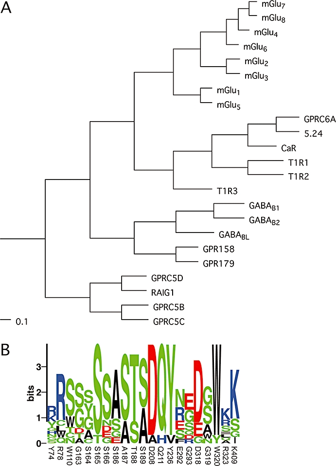
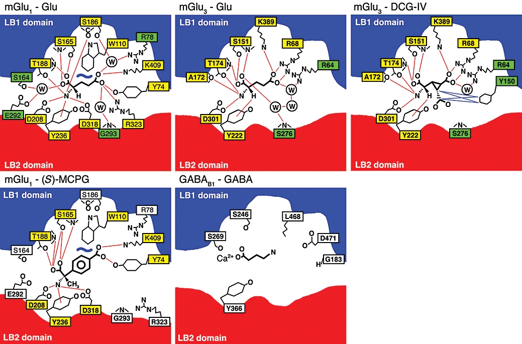
Family C of human G-protein-coupled receptors (GPCRs) is constituted by eight metabotropic glutamate receptors, two gamma-aminobutyric acid type B (GABA(B1-2)) subunits forming the heterodimeric GABA(B) receptor, the calcium-sensing receptor, three taste1 receptors (T1R1-3), a promiscuous L-alpha;-amino acid receptor G-protein-coupled receptor family C, group 6, subtype A (GPRC6A) and seven orphan receptors. Aside from the orphan receptors, the family C GPCRs are dimeric receptors characterized by a large extracellular Venus flytrap domain which bind the endogenous agonists. Except from the GABA(B1-2) and T1R2-3 receptor, all receptors are either activated or positively modulated by amino acids. In this review, we outline mutational, biophysical and structural studies which have elucidated the interaction of the amino acids with the Venus flytrap domains, molecular mechanisms of receptor selectivity and the initial steps in receptor activation.


Figures

Figure 1
Figure 1
(A) Phylogenetic analysis of family C GPCRs based on their seven transmembrane domains. A multiple sequence alignment of the predicted seven transmembrane domains was generated using the program ClustalX 2.0.9, and the shown phylograms were generated using the unweighted pair group method with arithmetic mean (UPGMA) algorithm and viewed with the program TreeviewX 0.5.0. The scale bars are a function of amino acid substitutions based on the Gonnet series substitution matrix. (B) Sequence logo of binding pocket residues in family C receptors, displaying the degree of conservation of each amino acid. Residues submitted are identical to those listed in Table 1 and numbers refer to the residue numbers in mGlu1. The height of each symbol is proportional to its frequency and colour coded according to polarity. The logo was generated using the server at the Center for Biological Sequence Analysis (http://www.cbs.dtu.dk/~gorodkin/appl/plogo.html) (Schneider and Stephens, 1990; Gorodkin et al., 1997). CaR, calcium-sensing receptor; GABAB, γ-aminobutyric acid type B; GPCR, G-protein-coupled receptor; GPRC6A, G-protein coupled receptor family C, group 6, subtype A; mGlu, metabotropic glutamate; T1R, taste1 receptor.
Figure 2
Figure 2
Model of a dimeric family C GPCR in its open–open/resting (left) and open–closed/active (right) conformations. The two conformations are in equillibrium with each other and additional conformations (not shown). Agonists and antagonists will shift the equillibrium towards the active or resting conformation respectively. The localizations of the Venus flytrap (VFT) domain, cysteine-rich domain (CRD) and seven transmembrane domain (7TM) are indicated. The models were constructed with the program MacPyMol using coordinates from PDB files 1EWT (mGlu1 open–open/resting VFT), 1EWK (mGlu1 open–closed/active VFT), 2E4U (mGlu3 CRR) and 2R4S (β2-adrenergic receptor 7TM). GPCR, G-protein-coupled receptor; mGlu, metabotropic glutamate.
Figure 3
Figure 3
The orthosteric binding site in mGlu and GABAB receptors. Schematic drawings of the binding of agonists (Glu, DCG-IV or GABA) or antagonist [(S)-MCPG] to mGlu1, mGlu3 or GABAB1. The mGlu1 and mGlu3 drawings are based on X-ray crystallographic structures (Kunishima et al., 2000; Tsuchiya et al., 2002; Muto et al., 2007) whereas the GABAB drawing is based on molecular modelling and mutational data (Galvez et al., 1999; 2000a; Jensen et al., 2001a). Red and blue lines indicates hydrogen-bonding and van der Wahls contacts respectively. Yellow and green filled boxes indicates direct or indirect (via water) contacts respectively. Redrawn from the listed references. DCG-IV, (2S,2′R,3′R)-2-(2′,3′-dicarboxycyclopropyl)glycine; GABAB, γ-aminobutyric acid type B; mGlu, metabotropic glutamate; (S)-MCPG, (S)-(α)-methyl-4-carboxyphenylglycine.
Figure 4
Figure 4
L-Amino acid selectivity profiles at CaR, GPRC6A and the T1R1+T1R3 heterodimer. Amino acids are grouped according to side chain charge and polarity. Data have been normalized to allow for comparison of relative amino acid preferences. The profile for CaR was generated by normalizing reported EC50 values from seven amino acids (measured in human parathyroid cells in the presence of 2 mmol·L−1 Ca2+) to the response of L-Trp (set to 100%). Amino acids marked with asterices (*) were not included in the original study. The profile for GPRC6A is based on reported EC50 values from mouse GPRC6A measured in the presence of 1 mmol·L−1 Ca2+ and 1 mmol·L−1 Mg2+ (Christiansen et al., 2007) and here normalized to the L-Lys response (set to 100%). T1R1+T1R3 data originally in the form of ‘number of responsive cells’ measured in the presence of 2.5 mmol·L−1 IMP (Nelson et al., 2001) were converted to percentage normalized response by calculating the response relative to that of L-Cys (set to 100%). The symbol (#) denotes that L-Tyr was not tested at T1R1+T1R3 (due to insolubility). CaR, calcium-sensing receptor; GPRC6A, G-protein coupled receptor family C, group 6, subtype A; T1R, taste1 receptor.

References

    1. Abe H, Tateyama M, Kubo Y. Functional identification of Gd3+ binding site of metabotropic glutamate receptor 1a. FEBS Lett. 2003;545:233–238. - PubMed
    1. Acher FC, Bertrand H-O. Amino acid recognition by Venus flytrap domains is encoded in an 8-residue motif. Biopolymers. 2005;80:357–366. - PubMed
    1. Alexander SP, Mathie A, Peters JA. Guide to Receptors and Channels (GRAC) Br J Pharmacol. (3rd edn) 2008;153(Suppl. 2):S1–S209. - PMC - PubMed
    1. Bai M, Quinn S, Trivedi S, Kifor O, Pearce SHS, Pollak MR, et al. Expression and characterization of inactivating and activating mutations in the human Ca2+o-sensing receptor. J Biol Chem. 1996;271:19537–19545. - PubMed
    1. Bai M, Trivedi S, Kifor O, Quinn SJ, Brown EM. Intermolecular interactions between dimeric calcium-sensing receptor monomers are important for its normal function. Proc Natl Acad Sci USA. 1999;96:2834–2839. - PMC - PubMed

Publication types

MeSH terms