. 2009 May;46(3):578-92.
doi: 10.1111/j.1469-8986.2009.00787.x. Epub 2009 Mar 4.

Validation of regression-based myogenic correction techniques for scalp and source-localized EEG

Affiliations

Validation of regression-based myogenic correction techniques for scalp and source-localized EEG

Brenton W McMenamin et al. Psychophysiology. 2009 May.

Abstract

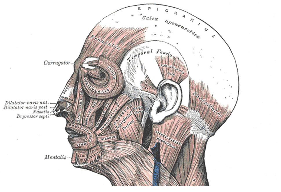
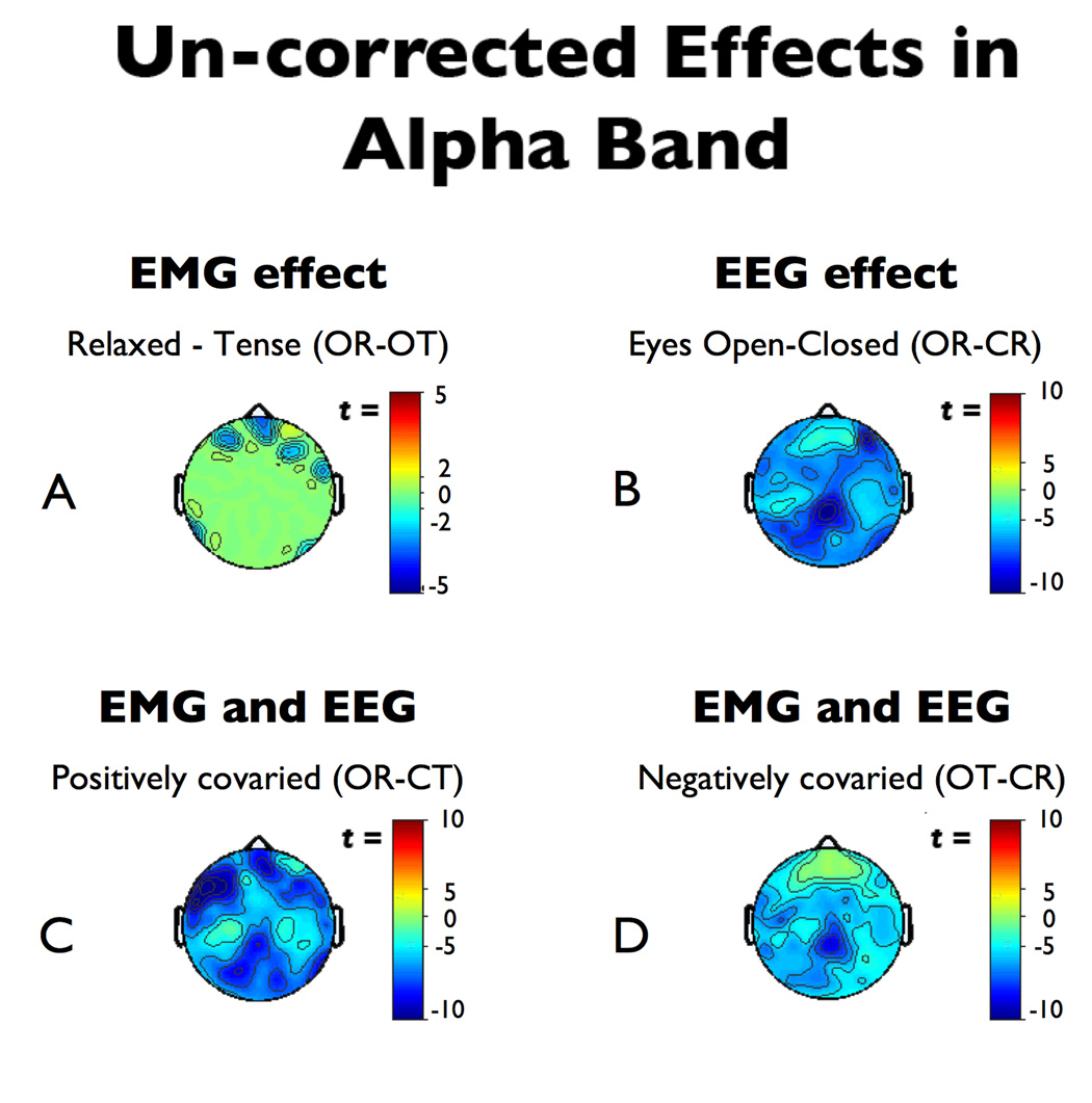
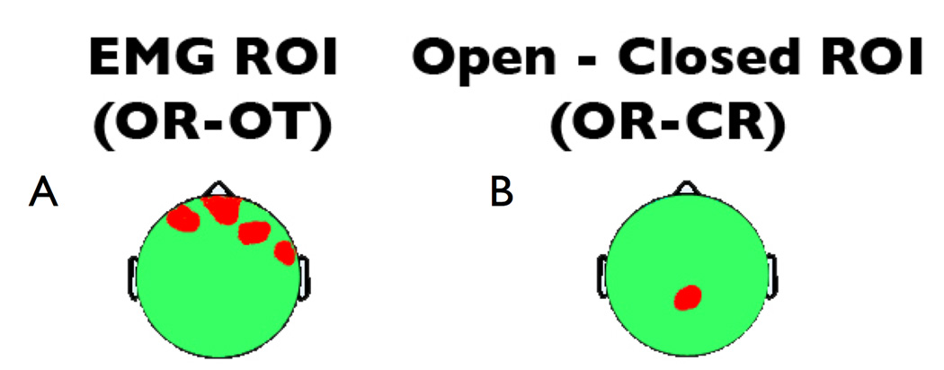
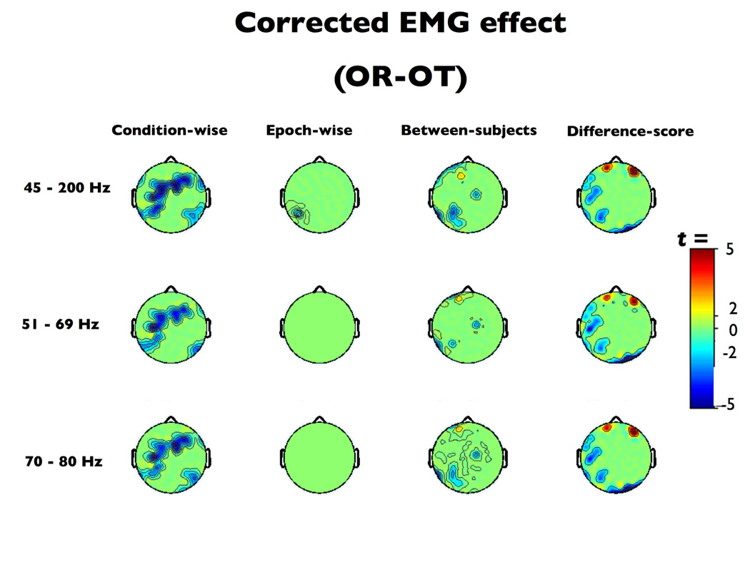
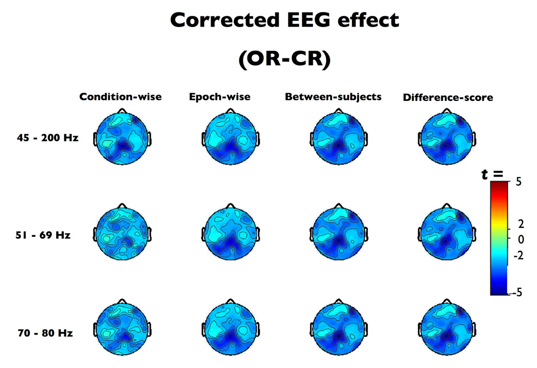
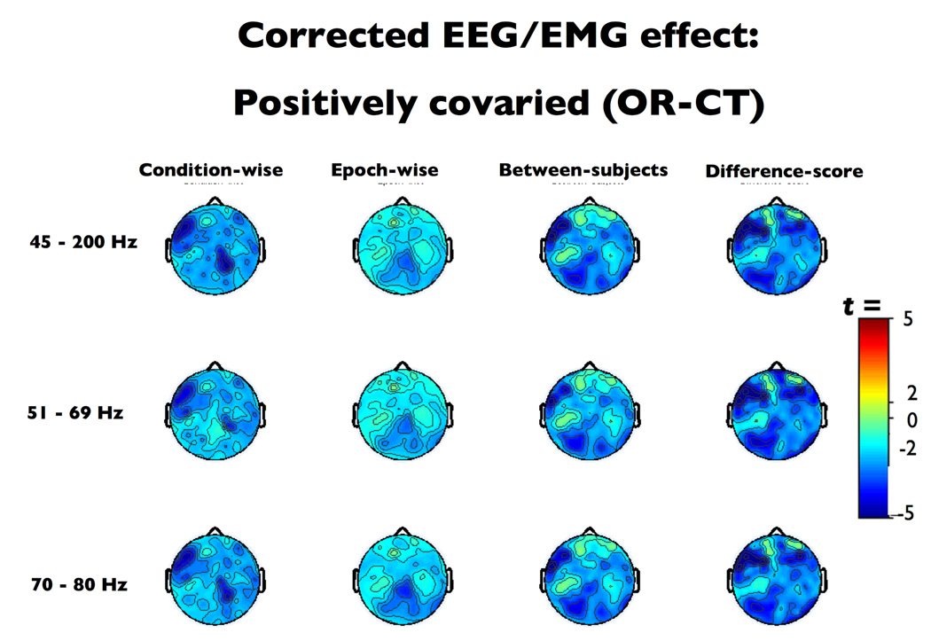
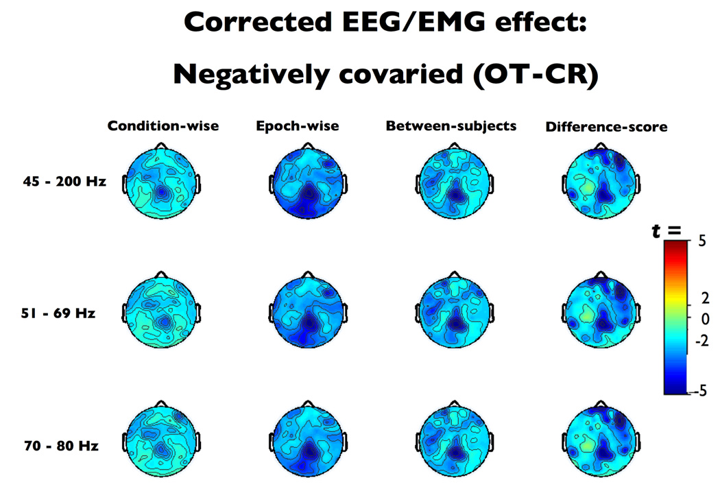
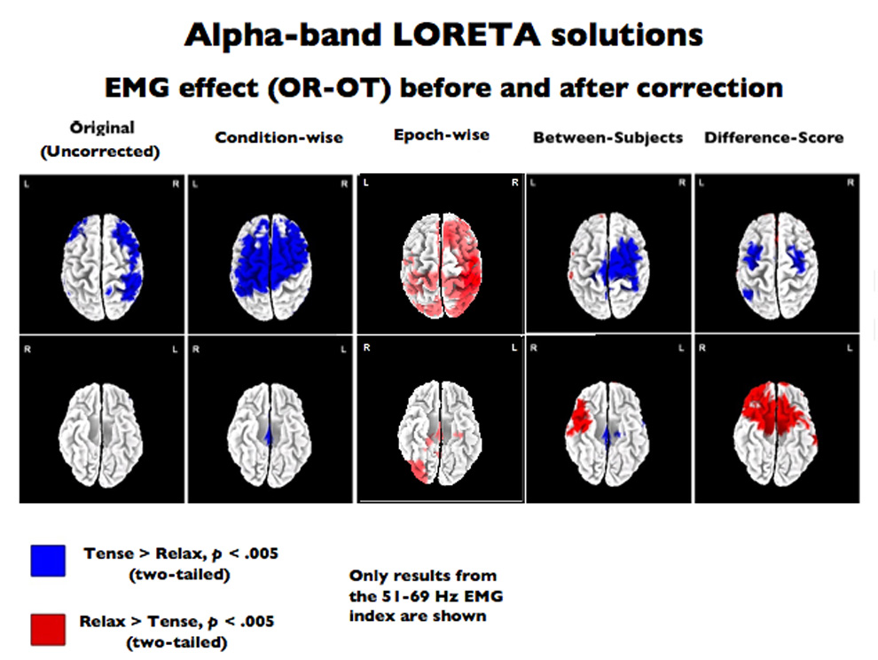
EEG and EEG source-estimation are susceptible to electromyographic artifacts (EMG) generated by the cranial muscles. EMG can mask genuine effects or masquerade as a legitimate effect-even in low frequencies, such as alpha (8-13 Hz). Although regression-based correction has been used previously, only cursory attempts at validation exist, and the utility for source-localized data is unknown. To address this, EEG was recorded from 17 participants while neurogenic and myogenic activity were factorially varied. We assessed the sensitivity and specificity of four regression-based techniques: between-subjects, between-subjects using difference-scores, within-subjects condition-wise, and within-subject epoch-wise on the scalp and in data modeled using the LORETA algorithm. Although within-subject epoch-wise showed superior performance on the scalp, no technique succeeded in the source-space. Aside from validating the novel epoch-wise methods on the scalp, we highlight methods requiring further development.


Figures

Figure 1
Figure 1. Illustration of the cranial musculature
The cranial musculature as depicted in Gray’s Anatomy (Gray, 1918; http://www.bartleby.com/). Nearly all cephalic electrodes lie on top of or adjacent to muscle, making EMG contamination a concern for the entire head – not just anterior electrodes.
Figure 2
Figure 2. Uncorrected effects in the alpha-band
Thresholded (p < .01) effects in the alpha-band (8–13 Hz) prior to EMG-correction. A) The purely myogenic effect (OR-OT). Note the significant myogenic activation near the mastoids. B) The alpha-blocking neurogenic effect (OR-CR). Note the roughly two-fold larger effect-size compared to the purely myogenic effect. C) The effect of positively covaried EMG and alpha-blocking (OR-CT). Note the shift in the peak from the posterior midline to the left anterior region. D) The effect of negatively covaried EMG and alpha-blocking (OT-CR). Abbreviations are described in the Method section.
Figure 3
Figure 3. Regions of interest used for analyses of sensitivity and specificity
Regions of interest (ROIs) used to quantify the sensitivity and specificity of EMG correction on the scalp. a) the Myogenic/EMG ROI (OR-OT contrast) used to assess sensitivity of EMG correction and b) the Neurogenic/EEG ROI (OR-CR contrast) used to assess specificity of EMG correction.
Figure 4
Figure 4. The eyes open relaxed-tense contrast after EMG-residualization
Thresholded (p < .01) t-scores for the eyes open relaxed-tense (OR-OT) contrast after EMG-residualization. Note the relatively poor performance of the condition-wise method.
Figure 5
Figure 5. The relaxed eyes open-closed contrast after EMG-residualization
Thresholded (p < .01) t-scores for eyes open-closed, relaxed (OR-CR) contrast after EMG-residualization.
Figure 6
Figure 6. The eyes open, relaxed minus eyes closed, tense contrast after EMG-residualization
Thresholded (p < .01) t-scores for the positively confounded eyes open, relaxed – closed, tense (OR–CT) contrast after EMG-residualization. At a qualitative level, only the epoch-wise method recovered the topography of the neurogenic effect (cf. Figure 2b).
Figure 7
Figure 7. The eyes open, tense minus eyes closed, relaxed contrast after EMG-residualization
Thresholded (p < .01) t-scores for the negatively confounded eyes open, tense – closed, relaxed (OT–CR) contrast after EMG-residualization. Note the poor sensitivity of the condition-wise technique.
Figure 8
Figure 8. The eyes open relaxed-tense (i.e., purely myogenic) contrast after EMG-residualization on LORETA data
LORETA results. Thresholded (p < .005, uncorrected) eyes open relaxed-tense (OR-OT) contrast in the LORETA source-space rendered on the dorsal (top) and ventral (bottom) surfaces of a canonical brain. The left-most image depicts the uncorrected contrast (i.e., prior to EMG-correction), followed by depictions (from left to right) of the effect after residualizing with the condition-wise, epoch-wise, between- and difference-score methods using the 51–69Hz EMG-band. Results (not shown) were similar for the other EMG-bands.

References

    1. Allen JJ, Coan JA, Nazarian M. Issues and assumptions on the road from raw signals to metrics of frontal EEG asymmetry in emotion. Biological psychology. 2004;67(1–2):183–218. - PubMed
    1. Bai X, Towle VL, He EJ, He B. Evaluation of cortical current density imaging methods using intracranial electrocorticograms and functional MRI. Neuroimage. 2007;35(2):598–608. - PMC - PubMed
    1. Barlow JS. EMG artifact minimization during clinical EEG recordings by special analog filtering. Electroencephalography and clinical neurophysiology. 1984;58(2):161–174. - PubMed
    1. Berg P, Scherg M. Dipole models of eye movements and blinks. Electroencephalography and clinical neurophysiology. 1991;79(1):36–44. - PubMed
    1. Berger H. Uber das elektroenkephalogramm des Menschen IV. Archiv fur Psychiatrie und Nervenkrankheiten. 1932;97:6–26.

Publication types

LinkOut - more resources