. 2010 Jan;20(1):106-18.
doi: 10.1111/j.1750-3639.2008.00246.x. Epub 2009 Mar 3.

Lithium restores neurogenesis in the subventricular zone of the Ts65Dn mouse, a model for Down syndrome

Affiliations

Lithium restores neurogenesis in the subventricular zone of the Ts65Dn mouse, a model for Down syndrome

Patrizia Bianchi et al. Brain Pathol. 2010 Jan.

Abstract

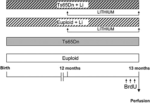
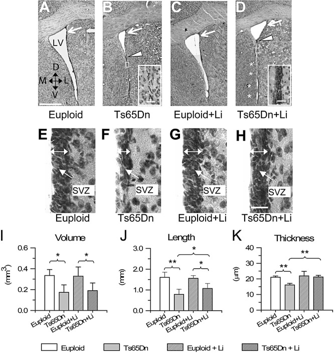
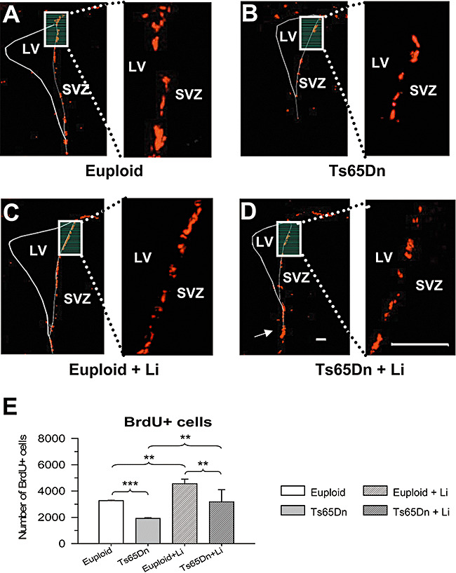
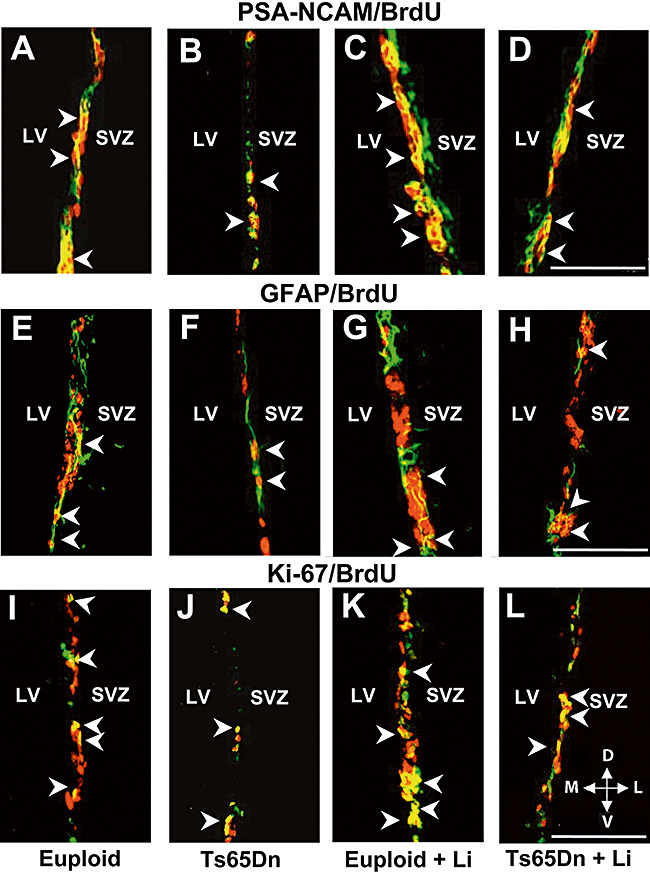
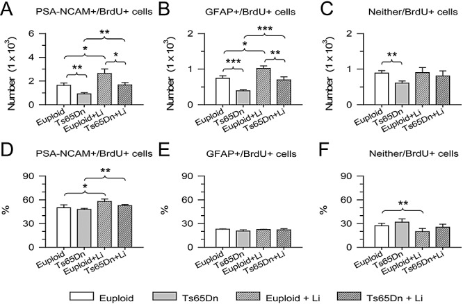
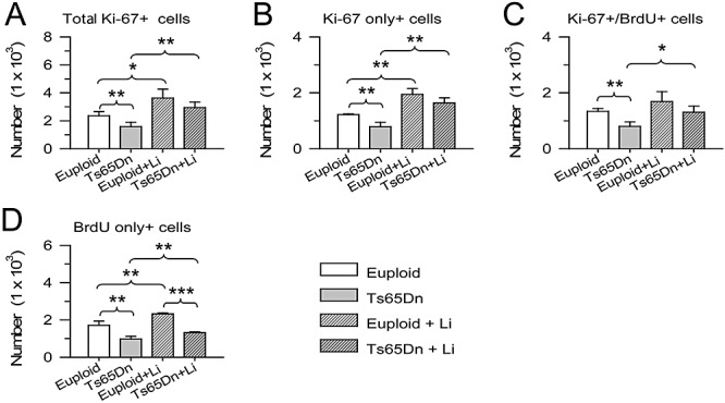
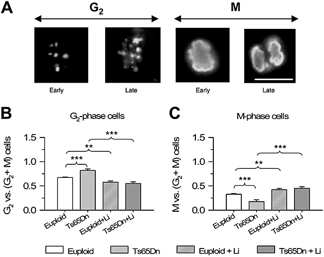
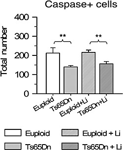
Down syndrome (DS), a high-incidence genetic pathology, involves brain hypoplasia and mental retardation. Emerging evidence suggests that reduced neurogenesis may be a major determinant of brain underdevelopment in DS. To establish whether it is possible to improve neurogenesis in DS, Ts65Dn mice--the most widely used model for DS--and euploid mice were treated with control or lithium chow for 1 month. During the last 3 days animals received one daily injection of 5-bromo-2-deoxyuridine (BrdU)--a marker of proliferating cells--and were sacrificed 24 h after the last injection. Neurogenesis was examined in the subventricular zone (SVZ), a region that retains a neurogenic potential across life. We found that Ts65Dn mice had less (-40%) BrdU+ cells than euploid mice, indicating severe proliferation impairment. Treatment with lithium increased the number of Brdu+ cells in both euploid and Ts65Dn mice. In the latter the number of Brdu+ cells became similar to that of untreated euploid mice. Our study shows that lithium is able to restore cell proliferation in the SVZ of the Ts65Dn mouse and point at treatments with mood stabilizers as a potential tool to improve neurogenesis in patients with DS.


Figures

Figure 1
Figure 1
Experimental protocol. Starting from the age of 12 months, euploid (n = 3) and Ts65Dn (n = 3) mice were treated with lithium, administered through the food pellets for 1 month. Age‐matched euploid (n = 3) at Ts65Dn (n = 3) animals were used as controls. During the last 3 days animals of all groups received a daily intraperitoneal injection (150 µg/g body weight) of BrdU and were sacrificed 24 h after the last BrdU injection.
Figure 2
Figure 2
A–E. Pattern of the lateral ventricle and subventricular zone (SVZ). Examples of Nissl‐stained sections at the level of the rostral portion of the lateral ventricle (LV) in an untreated euploid mouse (A), an untreated Ts65Dn mouse (B), a euploid mouse treated with lithium (C) and a Ts65Dn mouse treated with lithium (D). The plane of the section roughly corresponds to plane +0.50 of Franklin and Paxinos atlas (16). Note that the lumen of the ventricle is largely obliterated in the Ts65Dn mouse (region below the arrowhead). The insets in B and D show a portion of the ventricle where the lumen is obliterated (region enclosed by the rectangle) at a higher magnification. Note the absence of a SVZ in the untreated Ts65Dn mouse (B) and the appearance of a SVZ after treatment with lithium. The asterisks in D indicate small apertures in the obliterated ventricle. E–H. Higher magnification images of the lateral wall of the LV, at the level indicated by the white arrows in A–D in an untreated euploid mouse (E), an untreated Ts65Dn mouse (F), a euploid mouse treated with lithium (G) and a Ts65Dn mouse treated with lithium (H). The double‐headed arrows indicate the SVZ. Calibration in A = 500 µm, applies to A–D. Calibration in the insets in B,D = 25 µm. Calibration in H = 20 µm, applies to E–H. I–K. Stereology of the LV and SVZ. Volume of the LV (I), length of the lateral wall of the ventricle (J) and thickness of the SVZ (K) in untreated euploid and Ts65Dn mice, and in euploid and Ts65Dn mice treated with lithium. *P < 0.05; **P < 0.01 (Duncan's test after analysis of variance). Abbreviations: D = dorsal; L = lateral; M = medial; V = ventral.
Figure 3
Figure 3
Effect of lithium on cell proliferation in the subventricular zone (SVZ) of euploid and Ts65Dn mice. A–D. Examples of sections immunostained for BrdU in an untreated euploid mouse (A), an untreated Ts65Dn mouse (B), a euploid mouse treated with lithium (C) and a Ts65Dn mouse treated with lithium (D). The white rectangle in A–D indicates the region shown at a higher magnification on the right. Brdu+ cells (red) are mainly present along the lateral wall of the ventricle. Note that in the Ts65Dn mouse treated with lithium, BrdU+ cells were present also in the region where the lateral ventricle (LV) was obliterated (D, arrow). Calibrations in D apply to A–D: calibration of low magnification images = 200 µm; calibration of high magnification images = 120 µm. E. Number of BrdU+ cells in untreated euploid and Ts65Dn mice and euploid and Ts65Dn mice treated with lithium. **P < 0.01; ***P < 0.001 (Duncan's test after analysis of variance).
Figure 4
Figure 4
Double fluorescence immunohistochemistry. A–D: Examples of BrdU+ cells (red), PSA‐NCAM+ cells (green) and cells that are co‐labeled with BrdU and PSA‐NCAM (yellow) in the subventricular zone (SVZ) of a euploid mouse (A), a Ts65Dn mouse (B), a euploid mouse treated with lithium (C) and a Ts65Dn mouse treated with lithium (D). The arrowheads indicate some of the double‐labeled cells. E–H. Examples of BrdU+ cells (red), GFAP+ cells (green) and cells that are co‐labeled with BrdU and GFAP (yellow) in the SVZ of a euploid mouse (E), a Ts65Dn mouse (F), a euploid mouse treated with lithium (G) and a Ts65Dn mouse treated with lithium (H). The arrowheads indicate some of the double‐labeled cells. I–L. Examples of BrdU+ cells (red), Ki‐67+ cells (green) and cells that are co‐labeled with BrdU and Ki‐67 (yellow) in the SVZ of a euploid mouse (I), a Ts65Dn mouse (J), a euploid mouse treated with lithium (K) and a Ts65Dn mouse treated with lithium (L). The arrowheads indicate some of the double‐labeled cells. Calibration = 75 µm. Abbreviations: D = dorsal; GFAP = glial fibrillary acidic protein; L = lateral; LV = lateral ventricle; M = medial; PSA‐NCAM = polysialylated neural cell adhesion molecule; V = ventral.
Figure 5
Figure 5
Phenotype of the proliferating cells in the subventricular zone (SVZ). Total number (A) and percentage (D) of cells co‐labeled with BrdU and PSA‐NCAM, total number (B) and percentage (E) of cells co‐labeled with BrdU and PSA‐NCAM, and total number (C) and percentage (F) of cells that did not express neither PSA‐NCAM nor GFAP. Cells expressing PSA‐NCAM correspond to type A cells, cells expressing GFAP correspond to type B2 and cells that do not express either marker are type C cells. *P < 0.05; **P < 0.01; ***P < 0.001 (Duncan's test after analysis of variance). Abbreviations: GFAP = glial fibrillary acidic protein; PSA‐NCAM = polysialylated neural cell adhesion molecule.
Figure 6
Figure 6
Effect of lithium on the size of the proliferating population in the subventricular zone of euploid and Ts65Dn mice. A. Total number of Ki‐67+ cells. B. Number of Ki‐67+ cells that were not co‐labeled with BrdU. C. Number of Ki‐67+ cells that were co‐labeled with BrdU. D. Number of BrdU+ cells that were not co‐labeled with Ki‐67. *P < 0.05; **P < 0.01; ***P < 0.001 (Duncan's test after analysis of variance).
Figure 7
Figure 7
Effect of lithium on the percentage of cells in G2 and M phases of the cell cycle. A. Images of cells positive for phosphorylated‐histone‐H3 (pHH3) in the subventricular zone of a euploid mouse, showing a cell in early and late G2, early and late M phase. Cells in G2 exhibit a discontinuous pHH3 nuclear staining, cells in the early M phase exhibit a homogeneously condensed pattern and cells approaching division (late M phase) exhibit mitotic spindles. Scale bar (18 µm) applies to all the figures. B,C. Percentage of cells in G2 (B) and M (C) phases of the cell cycle with respect to total number of pHH3‐positive cells. **P < 0.01; ***P < 0.001 (Duncan's test after analysis of variance).
Figure 8
Figure 8
Effect of lithium on apoptotic cell death in euploid and Ts65Dn mice. Total number of cleaved caspase‐3‐positive cells in the subventricular zone of animals untreated and treated with lithium. **P < 0.01 (Duncan's test after analysis of variance).

References

    1. Aylward EH, Habbak R, Warren AC, Pulsifer MB, Barta PE, Jerram M, Pearlson GD (1997) Cerebellar volume in adults with Down syndrome. Arch Neurol 54:209–212. - PubMed
    1. Aylward EH, Li Q, Honeycutt NA, Warren AC, Pulsifer MB, Barta PE, Chan MD (1999) MRI volumes of the hippocampus and amygdala in adults with Down's syndrome with and without dementia. Am J Psychiatry 156:564–568. - PubMed
    1. Berridge MJ, Downes CP, Hanley MR (1989) Neural and developmental actions of lithium: a unifying hypothesis. Cell 59:411–419. - PubMed
    1. Blomgren K, Leist M, Groc L (2007) Pathological apoptosis in the developing brain. Apoptosis 12:993–1010. - PubMed
    1. Brazel CY, Romanko MJ, Rothstein RP, Levison SW (2003) Roles of the mammalian subventricular zone in brain development. Prog Neurobiol 69:49–69. - PubMed

Publication types

MeSH terms